Tracheoesophageal fistula (TEF) is a congenital condition characterized by an abnormal connection between the trachea and the esophagus, often leading to severe feeding difficulties, respiratory complications, and recurrent infections in newborns. Understanding the embryological basis of tracheoesophageal fistula is critical for clinicians, surgeons, and researchers in order to provide accurate diagnosis, plan surgical repair, and anticipate potential associated anomalies. The development of the respiratory and digestive tracts is a complex process that involves precise interactions between endodermal and mesodermal tissues. Disruptions in these processes can lead to the formation of TEF, which is frequently observed in combination with esophageal atresia.
Normal Embryological Development of the Foregut
During early embryogenesis, the foregut gives rise to both the respiratory and digestive systems. By the fourth week of gestation, a ventral outpouching known as the laryngotracheal diverticulum forms from the anterior wall of the primitive foregut. This diverticulum elongates and eventually separates from the dorsal foregut to form the trachea and the lung buds, while the dorsal foregut develops into the esophagus. The process of separation is orchestrated by the formation of the tracheoesophageal septum, a mesodermal structure that grows to divide the ventral and dorsal components of the foregut. Proper development of this septum is crucial for ensuring that the trachea and esophagus become distinct structures.
Formation of the Tracheoesophageal Septum
The tracheoesophageal septum forms through a combination of epithelial proliferation, apoptosis, and mesenchymal remodeling. Initially, the foregut is a single tube lined by endoderm and surrounded by splanchnic mesoderm. The lateral walls of the foregut thicken and fold medially, creating ridges that eventually fuse to form the septum. Disruption in the timing, migration, or fusion of these ridges can prevent complete separation of the trachea and esophagus, leading to a persistent fistulous connection. This developmental failure explains the diverse anatomical presentations of TEF, which can range from isolated fistulas to more complex forms associated with esophageal atresia.
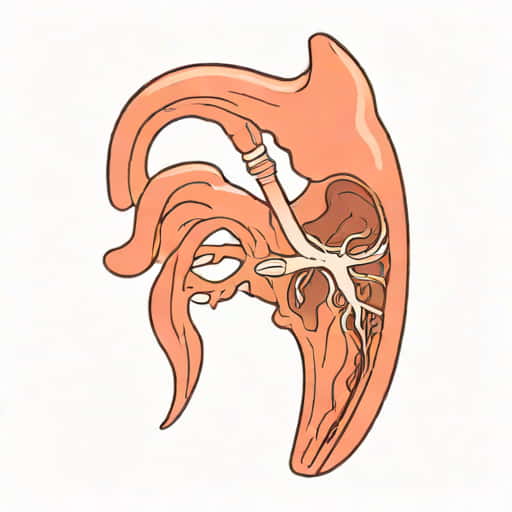
Types of Tracheoesophageal Fistula
Embryological anomalies in foregut separation result in several anatomical types of tracheoesophageal fistula, with the most common being the proximal esophageal atresia with distal TEF. Other types include isolated esophageal atresia without fistula, H-type fistulas where both trachea and esophagus are patent, and less frequent variants with long-segment fistulas. Each type reflects a specific pattern of failure in the tracheoesophageal septation process during early embryonic development.
- Proximal esophageal atresia with distal TEFThe most common form, occurring when the tracheoesophageal septum fails to close near the distal foregut.
- Isolated esophageal atresiaComplete separation without any fistulous connection, reflecting early failure in elongation of the esophagus.
- H-type fistulaBoth trachea and esophagus remain continuous, but an abnormal connection persists laterally, often difficult to detect clinically.
- Long-segment TEFRare forms with extensive fistulous tracts, resulting from broader mesodermal or endodermal defects.
Molecular and Genetic Influences
The embryological development of the trachea and esophagus is regulated by a complex network of signaling pathways, including Sonic Hedgehog (SHH), fibroblast growth factors (FGFs), Bone Morphogenetic Proteins (BMPs), and Wnt signaling. Alterations in these pathways can interfere with proper foregut separation, leading to tracheoesophageal malformations. For instance, mutations in the SHH pathway have been linked to both isolated and syndromic TEF cases, highlighting the importance of molecular control in the formation of the tracheoesophageal septum. Genetic studies have also identified familial clustering of TEF, suggesting heritable predisposition in certain populations.
Associated Syndromes and Anomalies
Tracheoesophageal fistula often occurs in association with other congenital anomalies, particularly those affecting the cardiac, renal, and vertebral systems. This association is commonly described as part of the VACTERL spectrum (Vertebral anomalies, Anal atresia, Cardiac defects, Tracheoesophageal fistula, Renal anomalies, Limb abnormalities). The embryological basis for these associations lies in the shared developmental timing and signaling pathways among these organ systems. Disruptions affecting the tracheoesophageal septum may concurrently impact the morphogenesis of nearby structures, explaining the frequent co-occurrence of TEF with other congenital defects.
Pathophysiology and Clinical Implications
Understanding the embryological origin of TEF helps explain the clinical challenges associated with this condition. Newborns with tracheoesophageal fistula may present with choking, coughing, cyanosis, and excessive salivation during feeding. The abnormal communication allows gastric contents or saliva to enter the trachea, leading to aspiration and recurrent respiratory infections. In addition, esophageal atresia often results in feeding obstruction and growth delays. The specific type of TEF, determined by the nature and location of the fistula, guides surgical planning and anticipates potential complications.
Surgical Considerations
Surgical repair of TEF aims to close the abnormal communication and restore normal continuity of the esophagus. A thorough understanding of the embryological development is essential for surgeons, as it informs the location of the fistula, the potential presence of a proximal pouch, and the risk of associated anomalies. Techniques often involve mobilization of the esophageal segments, ligation of the fistula, and careful preservation of surrounding structures to minimize post-operative complications. Early recognition and intervention improve outcomes, particularly in neonates with complex TEF or associated anomalies.
Research and Future Directions
Ongoing research into the embryological basis of tracheoesophageal fistula seeks to clarify the molecular mechanisms that drive foregut development. Studies using animal models, genetic analysis, and molecular biology techniques aim to identify key genes and signaling pathways that, when disrupted, result in TEF. Advances in prenatal imaging and molecular diagnostics hold promise for early detection and potential preventive strategies. Understanding the precise embryological timing and interactions involved in tracheoesophageal septation may also lead to novel therapeutic approaches and improved surgical outcomes.
Tracheoesophageal fistula arises from complex disruptions in the embryological development of the foregut, particularly involving the formation of the tracheoesophageal septum. Its diverse anatomical presentations reflect varying degrees of failure in foregut separation, influenced by genetic, molecular, and environmental factors. Recognizing the embryological basis of TEF not only enhances our understanding of its pathophysiology but also informs clinical management, surgical planning, and long-term care. As research continues to elucidate the molecular mechanisms underlying foregut development, there is potential for improved diagnostic tools, preventive strategies, and therapeutic interventions, ultimately improving outcomes for affected neonates and their families.