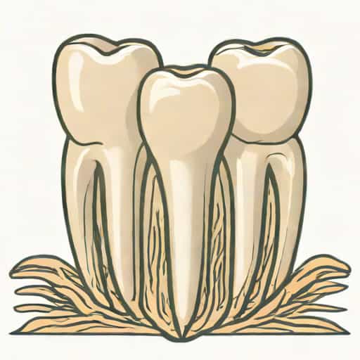
The maxillary second premolar is an important tooth in dentistry due to its unique anatomy, function, and variability in root morphology. Understanding the number of roots in this tooth is essential for dental professionals, particularly for procedures such as root canal treatment, extractions, and restorative dentistry. Unlike some other teeth, the maxillary second premolar exhibits a degree of variation in root number, which can affect clinical decision-making. A clear grasp of its anatomical features, including the number and configuration of roots, is critical for ensuring successful treatment outcomes and minimizing complications during dental procedures.
Anatomy of the Maxillary Second Premolar
The maxillary second premolar is located in the upper jaw, posterior to the first premolar and anterior to the first molar. It serves as a transitional tooth between the canine and molar teeth, combining features of both in terms of morphology and function. Its primary role includes aiding in the tearing and grinding of food and maintaining the alignment of the dental arch. The crown of the maxillary second premolar is typically oval or slightly rounded, with two cusps-buccal and lingual-that vary in size depending on the individual.
Root Morphology
One of the key characteristics of the maxillary second premolar is the number of roots it possesses. The root structure is essential for anchoring the tooth in the jawbone and providing stability during chewing. Most maxillary second premolars have a single root, but variations can occur, with two roots being observed in some cases. Understanding these variations is crucial for clinicians, especially when performing endodontic procedures where accurate identification of all roots is necessary to avoid treatment failure.
Number of Roots in Maxillary Second Premolars
Studies have shown that the majority of maxillary second premolars have a single root, making them relatively straightforward to treat in most dental procedures. However, a smaller percentage of these teeth may present with two roots, usually consisting of a buccal and a palatal root. Rarely, three-rooted maxillary second premolars have been documented, though this occurrence is extremely uncommon. Knowledge of these anatomical variations helps dental professionals anticipate potential challenges during treatment.
Single Root Configuration
The single root configuration is the most common, occurring in approximately 60 to 90 percent of maxillary second premolars, depending on population studies. This root usually has one or occasionally two canals, which are typically straight or slightly curved. Single-rooted teeth generally facilitate easier access during endodontic procedures, as the canals are easier to locate and negotiate. Nonetheless, clinicians must be aware of the possibility of additional canals within a single root to prevent missed anatomy and treatment failure.
Two Root Configuration
Approximately 10 to 30 percent of maxillary second premolars exhibit two roots, with one located buccally and the other palatally. This variation is more commonly observed in certain populations and requires careful radiographic assessment for accurate diagnosis. Two-rooted premolars often have two or more canals, which can be challenging to locate and clean thoroughly. Awareness of this possibility is critical for endodontic success, as missing a canal in a multi-rooted tooth is a common cause of persistent infection or post-treatment complications.
Rare Three-Rooted Premolars
Although extremely rare, three-rooted maxillary second premolars have been reported in case studies. These teeth usually have a configuration similar to maxillary molars, with two buccal roots and one palatal root. Identifying a three-rooted premolar requires careful clinical examination and advanced imaging techniques, such as cone-beam computed tomography (CBCT). Recognizing this rare variation is important for preventing procedural errors and ensuring comprehensive endodontic treatment.
Clinical Significance of Root Variations
Variations in root number and morphology of the maxillary second premolar have significant clinical implications. Accurate identification of root configuration influences the approach to endodontic treatment, extractions, orthodontic procedures, and restorative dentistry. Failure to recognize anatomical variations can lead to incomplete treatment, persistent infection, or structural complications. Therefore, dental professionals must be vigilant in assessing each tooth individually.
Endodontic Considerations
Root canal treatment is particularly impacted by root variations. Single-rooted premolars with a single canal are generally easier to treat, while two-rooted or three-rooted premolars require more careful evaluation. Clinicians must use diagnostic tools, such as periapical radiographs from multiple angles or CBCT scans, to detect additional roots or canals. Locating all canals, cleaning them thoroughly, and achieving proper obturation are essential for successful endodontic outcomes.
Extraction and Surgical Implications
The number of roots also affects tooth extraction and surgical planning. Single-rooted premolars are usually easier to extract, whereas multi-rooted premolars may require sectioning or more complex techniques to avoid root fracture and damage to surrounding tissues. Awareness of root variations helps in selecting the appropriate instruments and techniques, minimizing patient discomfort and reducing the risk of complications.
Orthodontic and Restorative Applications
Root anatomy is also important for orthodontic treatment planning and restorative procedures. The presence of multiple roots affects tooth movement, anchorage, and stability. In restorative dentistry, understanding root morphology ensures proper crown placement and prevents overloading of roots, which can compromise tooth longevity. Clinicians must consider these factors when designing treatment plans for maxillary second premolars.
Methods for Identifying Root Number
Identifying the number of roots in maxillary second premolars requires a combination of clinical examination and radiographic imaging. Careful assessment allows for accurate diagnosis and successful treatment planning.
Radiographic Examination
Standard periapical radiographs are commonly used to assess root morphology. Taking multiple angled views helps detect additional roots or unusual canal configurations. In complex cases, CBCT imaging provides three-dimensional visualization of roots and canals, allowing for precise identification of anatomical variations. Radiographic evaluation is essential before performing endodontic or surgical procedures to avoid missing canals or roots.
Clinical Assessment
During dental procedures, clinicians can often identify variations in root morphology by careful exploration of the pulp chamber and canal system. Using magnification tools such as loupes or microscopes improves visibility, making it easier to detect accessory canals and unusual root configurations. Combining clinical observation with imaging provides the most accurate assessment of root anatomy.
The maxillary second premolar exhibits a range of root variations that are clinically significant. While the majority of these teeth have a single root, a notable percentage have two roots, and rare cases present with three roots. Understanding these variations is critical for dental procedures such as root canal treatment, extractions, orthodontic planning, and restorative dentistry. Accurate diagnosis through radiographic imaging and careful clinical assessment ensures successful treatment outcomes, minimizes complications, and enhances patient care. Knowledge of the number of roots and their configurations allows dental professionals to approach each maxillary second premolar with confidence, providing safe and effective treatment tailored to the individual anatomy of the tooth.