Renal calculi, more commonly known as kidney stones, are a frequent topic in anatomy and physiology because they combine structural details of the urinary system with pathological processes that affect normal function. Understanding the anatomy and physiology of renal calculi helps explain how these stones develop, how they impact the kidneys and urinary tract, and why certain individuals are more prone to them. This subject is often explored in medical presentations or renal calculi PPT notes for students and healthcare professionals, making it a useful resource for learning both the basics and the complications of stone formation.
Anatomy of the Kidneys and Urinary Tract
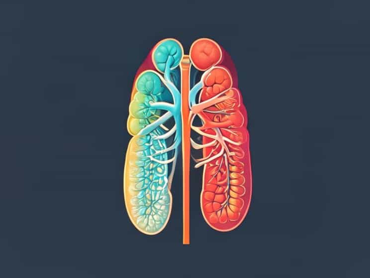
The kidneys are bean-shaped organs located in the retroperitoneal space of the abdomen. Their primary role is to filter blood, remove waste, and maintain fluid and electrolyte balance. A clear understanding of their anatomy is essential when discussing renal calculi.
Key Structures Involved
- Renal CortexThe outer layer of the kidney where filtration begins.
- Renal MedullaContains the renal pyramids, where urine is concentrated.
- CalycesCup-shaped structures that collect urine from the pyramids.
- Renal PelvisFunnel-shaped cavity that collects urine before passing it to the ureter.
- UretersTubes that carry urine from the renal pelvis to the bladder.
Renal calculi typically form in the calyces or renal pelvis and may move into the ureters, causing significant pain and urinary obstruction.
Physiology of Urine Formation
To understand renal calculi, one must first understand how urine is formed. The kidneys filter approximately 180 liters of blood plasma daily, but only about 1-2 liters of urine is excreted. This process involves three main steps
- FiltrationBlood plasma passes through the glomeruli into Bowman’s capsule.
- ReabsorptionEssential substances like glucose, electrolytes, and water are reabsorbed into the bloodstream.
- SecretionWaste products, hydrogen ions, and drugs are secreted into the filtrate.
Any imbalance in the concentration of solutes, hydration levels, or urinary pH can influence stone formation, highlighting the close relationship between renal physiology and calculi development.
What Are Renal Calculi?
Renal calculi are solid masses formed by crystallization of minerals and salts in the kidneys. They can vary in size from tiny grains to large stones that fill the renal pelvis. The chemical composition of stones often includes calcium oxalate, calcium phosphate, uric acid, or cystine.
Types of Renal Calculi
- Calcium StonesThe most common type, usually calcium oxalate or phosphate.
- Uric Acid StonesFormed in individuals with high uric acid levels or acidic urine.
- Struvite StonesOften associated with urinary tract infections and composed of magnesium, ammonium, and phosphate.
- Cystine StonesRare and caused by a hereditary disorder that leads to cystinuria.
Pathophysiology of Renal Calculi Formation
The development of renal calculi is a result of supersaturation of urine with stone-forming substances. When urine contains more crystal-forming substances than it can dilute, crystals start to stick together. Several factors contribute to this process
- SupersaturationExcess calcium, oxalate, uric acid, or cystine in the urine promotes crystallization.
- NucleationSmall ptopics act as a nucleus where crystals can form and grow.
- AggregationCrystals join together, forming larger stones.
- RetentionStones lodge in calyces or renal pelvis, resisting elimination.
In physiology, urine normally contains inhibitors such as citrate, which help prevent stone formation. A reduction in these inhibitors increases the risk of calculi.
Clinical Anatomy How Stones Cause Symptoms
The movement of stones from the renal pelvis to the ureter explains the hallmark symptoms of renal calculi. The ureter is narrow, so stones often get stuck, leading to intense pain known as renal colic.
Common Symptoms
- Severe flank pain that radiates to the lower abdomen or groin.
- Hematuria (blood in the urine) caused by stones scraping the urinary tract lining.
- Nausea and vomiting due to pain and autonomic stimulation.
- Frequent urge to urinate with small urine output.
- Possible fever and chills if infection is present.
Risk Factors for Renal Calculi
Both anatomy and physiology contribute to the risk of developing kidney stones. Factors include
- Dehydration leading to concentrated urine.
- Diet high in oxalate-rich foods like spinach and nuts.
- Excess salt intake, increasing calcium excretion.
- Genetic predisposition to stone formation.
- Urinary tract infections, which promote struvite stones.
- Metabolic disorders such as hyperparathyroidism or gout.
Diagnosis and Medical Relevance
Understanding the anatomy and physiology of renal calculi is essential for accurate diagnosis and treatment. Imaging techniques like ultrasound, X-rays, and CT scans are often used to locate stones. Laboratory analysis of urine and blood helps identify chemical imbalances contributing to stone formation.
Treatment Options
- HydrationDrinking plenty of fluids helps flush small stones.
- MedicationDrugs can manage pain, relax ureters, or reduce uric acid levels.
- Extracorporeal Shock Wave Lithotripsy (ESWL)Non-invasive method to break stones into smaller fragments.
- UreteroscopyEndoscopic removal of stones from the urinary tract.
- Surgical InterventionRequired for large or complex stones.
Prevention Through Physiology and Lifestyle
Preventing renal calculi is closely tied to understanding renal physiology. Maintaining balanced hydration and electrolyte levels supports normal urine flow and prevents supersaturation of solutes.
Preventive Measures
- Drink 2-3 liters of water daily to dilute urine.
- Reduce sodium intake to minimize calcium excretion.
- Limit consumption of oxalate-rich foods if prone to calcium oxalate stones.
- Maintain a balanced intake of calcium to reduce oxalate absorption.
- Monitor uric acid levels and follow dietary advice for gout-related stones.
The anatomy and physiology of renal calculi provide a framework for understanding how kidney stones form, how they impact the urinary system, and why treatment and prevention are essential. By examining renal structure, urine physiology, and the pathological mechanisms behind crystallization, healthcare providers and students gain a clearer picture of this common condition. Renal calculi not only highlight the complex interaction between anatomy and physiology but also emphasize the importance of lifestyle, hydration, and preventive care in maintaining kidney health.