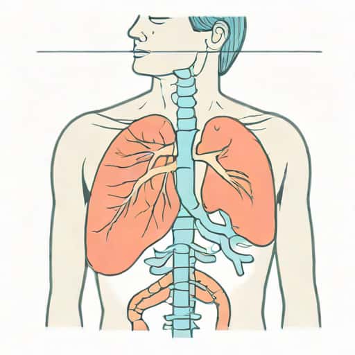
Tension pneumothorax is a life-threatening medical condition that occurs when air accumulates in the pleural space under pressure, leading to the collapse of the affected lung and significant impairment of cardiovascular function. One of the hallmark signs of this condition is jugular vein distention, which reflects the increased pressure in the thoracic cavity and the resulting difficulty in venous return to the heart. Recognizing tension pneumothorax early, understanding its relationship with jugular vein distention, and initiating rapid treatment are crucial for preventing severe complications and potentially fatal outcomes. This condition requires prompt medical attention and is considered a medical emergency in both trauma and non-trauma settings.
Understanding Tension Pneumothorax
Tension pneumothorax occurs when a one-way valve effect allows air to enter the pleural space during inspiration but prevents it from escaping during expiration. As air accumulates, intrathoracic pressure rises, compressing the lungs, heart, and major blood vessels. This compression can result in severe respiratory distress, hypoxia, hypotension, and shock. Unlike a simple pneumothorax, which may be managed conservatively in some cases, tension pneumothorax rapidly deteriorates if left untreated, making early recognition and intervention critical.
Causes and Risk Factors
Tension pneumothorax can develop from various causes, including trauma, medical procedures, or spontaneous events. Common causes include
- Blunt or penetrating chest trauma, such as motor vehicle accidents or stab wounds.
- Complications from mechanical ventilation or positive pressure ventilation in critically ill patients.
- Spontaneous pneumothorax, often in individuals with underlying lung disease such as chronic obstructive pulmonary disease (COPD) or cystic fibrosis.
- Rupture of pulmonary blebs or bullae, particularly in tall, thin, young adults.
- Medical procedures involving the thorax, such as central line placement, lung biopsy, or thoracentesis.
Jugular Vein Distention as a Clinical Sign
Jugular vein distention (JVD) is a visible bulging of the external jugular veins in the neck, often indicating increased central venous pressure. In the context of tension pneumothorax, JVD occurs because the rising intrathoracic pressure compresses the superior vena cava, impeding venous return to the heart. This obstruction leads to engorgement of the jugular veins and may be accompanied by hypotension, tachycardia, and cyanosis. Recognizing JVD as a sign of tension pneumothorax is essential for prompt diagnosis, especially in emergency and trauma settings.
Other Clinical Manifestations
In addition to jugular vein distention, tension pneumothorax presents with several critical signs and symptoms
- Severe shortness of breath and respiratory distress.
- Decreased or absent breath sounds on the affected side.
- Hyperresonance to percussion on the affected side of the chest.
- Tracheal deviation away from the affected side in advanced cases.
- Hypotension, tachycardia, and signs of shock.
- Cyanosis due to impaired oxygenation.
Pathophysiology Behind Jugular Vein Distention
The pathophysiology of jugular vein distention in tension pneumothorax involves increased intrathoracic pressure leading to compromised venous return. As air accumulates under pressure in the pleural space, it pushes mediastinal structures toward the opposite side, compressing the superior vena cava and right atrium. This compression prevents normal blood flow from the head and neck, causing the jugular veins to become visibly distended. Additionally, decreased cardiac output and impaired systemic perfusion contribute to hypotension and shock, further emphasizing the urgency of treatment.
Diagnosis
Rapid diagnosis of tension pneumothorax is primarily clinical, especially in emergency situations where imaging may not be immediately available. Key diagnostic steps include
- Physical examination noting respiratory distress, absent breath sounds, hyperresonance, tracheal deviation, and JVD.
- Assessment of vital signs, including hypotension, tachycardia, and hypoxia.
- Point-of-care ultrasound or chest X-ray, if available, to confirm lung collapse and mediastinal shift.
- Recognition of the clinical context, such as recent trauma, mechanical ventilation, or spontaneous events.
Emergency Management
Tension pneumothorax is a true medical emergency that requires immediate intervention to prevent cardiovascular collapse and death. The first-line treatment is needle decompression followed by chest tube insertion
Needle Decompression
Needle decompression is performed by inserting a large-bore needle or catheter into the second intercostal space at the midclavicular line on the affected side. This procedure allows trapped air to escape, relieving intrathoracic pressure and reducing compression of the heart and major vessels. Needle decompression is a temporizing measure and must be followed by definitive treatment with a chest tube.
Chest Tube Thoracostomy
After initial decompression, a chest tube is inserted into the pleural space to continuously evacuate air and allow the lung to re-expand. Chest tube thoracostomy is considered definitive management and stabilizes the patient while preventing recurrence of tension pneumothorax. Continuous monitoring of vital signs, oxygenation, and respiratory function is essential during and after the procedure.
Complications and Prognosis
If tension pneumothorax is not promptly treated, it can lead to severe complications, including cardiac arrest, irreversible hypoxia, and multi-organ failure. Jugular vein distention is a warning sign that indicates significant cardiovascular compromise. Early recognition and intervention significantly improve prognosis, and most patients recover fully with appropriate emergency care. However, delayed treatment can result in fatal outcomes, highlighting the importance of rapid assessment and immediate management in clinical practice.
Prevention and Risk Mitigation
In high-risk settings, such as trauma centers or intensive care units, prevention and early recognition of tension pneumothorax are critical. Strategies include
- Careful monitoring of patients on mechanical ventilation for signs of barotrauma.
- Prompt evaluation of chest trauma with clinical assessment and imaging.
- Education of healthcare professionals in rapid recognition of JVD and other signs of tension pneumothorax.
- Preparation of emergency equipment, including needles and chest tubes, for immediate intervention.
- Close observation of patients with known lung pathology for early symptoms of pneumothorax.
Tension pneumothorax is a medical emergency characterized by the accumulation of air in the pleural space under pressure, leading to lung collapse, cardiovascular compromise, and life-threatening consequences. Jugular vein distention serves as a critical clinical sign, reflecting increased intrathoracic pressure and impaired venous return to the heart. Early recognition, rapid needle decompression, and subsequent chest tube thoracostomy are essential for saving lives and preventing complications. Understanding the pathophysiology, clinical manifestations, and emergency management of tension pneumothorax is vital for healthcare providers, especially in trauma and critical care settings.
final remarkss
Awareness of tension pneumothorax and the significance of jugular vein distention can improve outcomes in emergency situations. Timely intervention, proper training, and vigilance in monitoring at-risk patients are essential components of effective management. By recognizing early warning signs, including JVD, and acting decisively, healthcare professionals can prevent fatal consequences and ensure optimal recovery for patients facing this potentially deadly condition.