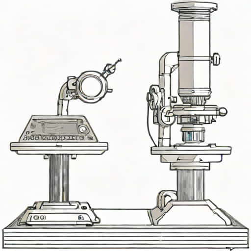
The resolution of an electron microscope measured in angstroms is a topic that fascinates researchers, students, and anyone intrigued by the hidden structures of matter. Understanding how this remarkable instrument achieves such extraordinary precision helps explain why it has become an essential tool in biology, materials science, and nanotechnology. By exploring how electron wavelengths, optical components, and instrument design contribute to the final resolution, it becomes easier to appreciate the science behind imaging at the atomic scale.
What Resolution in Electron Microscopy Really Means
Resolution refers to the ability of a microscope to distinguish two points as separate objects. The smaller the distance that can be resolved, the clearer and more detailed the image will be. In electron microscopy, this distance is often expressed in angstroms, a unit equal to one-tenth of a nanometer. Using angstrom measurements makes it possible to describe atomic-level structures with accuracy and clarity.
Why Angstroms Are Important
At the atomic and molecular scale, features become so small that conventional units such as micrometers lose their usefulness. Angstroms provide a more manageable scale for describing atomic spacing, electron shells, and lattice arrangements. Because many biological and material structures measure only a few angstroms across, the electron microscope’s ability to produce images at this scale is a major scientific advantage.
-
1 Å = 0.1 nanometers
-
Atoms typically range from 1 to 3 Å in diameter
-
Bonds between atoms often measure around 1 2 Å
The Role of Electron Wavelength in Achieving High Resolution
The most fundamental reason electron microscopes can achieve such extraordinary resolution is the extremely short wavelength of electrons when they are accelerated at high voltages. Light microscopes are limited by the wavelength of visible light, which ranges from 400 to 700 nanometers. In contrast, accelerated electrons can have wavelengths less than 0.05 angstroms, allowing the instrument to visualize structures far smaller than those visible under a light microscope.
How Acceleration Affects Wavelength
When electrons pass through an electric field, they gain energy and behave like waves with shorter wavelengths. The faster the electrons travel, the shorter the wavelength becomes, which directly improves resolution. This connection is based on the de Broglie equation, which describes how ptopics behave as waves.
-
Higher accelerating voltages result in shorter electron wavelengths
-
Shorter wavelengths lead to better theoretical resolution
-
Practical resolution is influenced by lens quality and system imperfections
Realistic Limits of Electron Wavelengths
Although the theoretical wavelength of electrons can be extremely small, real-world electron microscopes face practical limitations. Aberrations in the electromagnetic lenses, mechanical vibrations, contamination, and detector performance all influence the final resolution that can be achieved.
Types of Electron Microscopes and Their Angstrom-Level Resolution
Different types of electron microscopes provide different levels of resolution. Each type uses unique imaging systems and operational principles that determine its capabilities.
Transmission Electron Microscope (TEM)
The TEM is well-known for achieving some of the highest resolutions available in microscopy. Because it transmits electrons through an ultra-thin specimen, it can produce images that reveal atomic arrangements in materials or cellular structures in biology.
-
Typical resolution around 1 2 Å for advanced instruments
-
With aberration correction below 1 Å in some cases
-
Used for atomic-scale imaging, crystallography, and molecular analysis
Scanning Electron Microscope (SEM)
SEM is widely used for surface analysis. While it does not reach the same atomic-level resolution as TEM, it still produces extremely sharp images of surface structures.
-
Typical resolution 5 10 Å for high-end systems
-
Surface topology and morphology studies
Scanning Transmission Electron Microscope (STEM)
STEM combines aspects of both TEM and SEM. It scans a focused electron beam across a specimen and collects signals that vary with density and composition. With modern technology, STEM instruments can achieve sub-angstrom resolution, making them one of the most powerful tools for nanoscale imaging.
-
Resolution often reaches below 1 Å with aberration correction
-
Allows chemical analysis at atomic resolution
Aberration Correction and Its Impact on Angstrom Resolution
One of the most significant advancements in electron microscopy has been the development of aberration-corrected lenses. Historically, spherical and chromatic aberrations limited resolution, preventing microscopes from reaching the theoretical limit of electron wavelength. Aberration correctors use advanced electromagnetic fields to counteract distortions, significantly improving clarity.
Spherical Aberration Correction
Spherical aberration occurs because electrons passing through different parts of a lens converge at slightly different points. Correcting this distortion allows the electron beam to focus more sharply, dramatically enhancing resolution.
-
Reduced image blur
-
Improved contrast and definition
-
Atomic-level imaging becomes more reliable
Chromatic Aberration Correction
Chromatic aberration occurs when electrons with slightly different energies focus at different points. Although more challenging to correct, technologies continue to advance in minimizing this effect, bringing microscopes closer to theoretical resolution limits.
Factors That Influence Practical Resolution
Even with powerful technology, several external factors influence the actual resolution achieved during imaging. Researchers must control these elements carefully to obtain the highest quality results.
Sample Preparation
Specimens must be extremely thin for TEM and STEM imaging. Any irregularities, contamination, or thickness variations can reduce resolution.
-
Proper cleaning and preparation are essential
-
Freeze-drying and cryo-techniques help preserve delicate structures
Instrument Stability
Vibrations, electromagnetic interference, and temperature fluctuations can distort images. Modern electron microscopes are often placed in isolated rooms designed for maximum stability.
Vacuum Quality
A high vacuum is necessary to allow electrons to travel without scattering. Poor vacuum conditions can reduce resolution and cause additional noise in the image.
Applications of Angstrom-Level Resolution
Achieving resolution in angstroms allows scientists to visualize structures previously impossible to observe. This capability has transformed multiple scientific disciplines.
Materials Science
Researchers can examine crystal defects, atom spacing, and lattice structures. Understanding these features is essential for developing new materials, semiconductors, and nanotechnologies.
Biological Science
Although biological samples pose challenges due to sensitivity and the need for cryo techniques, angstrom-level imaging helps reveal protein structures, viruses, and membrane details.
Nanotechnology
Electron microscopes are crucial tools for designing and analyzing nanoscale devices, coatings, and molecular machines.
Seeing the Atomic World Clearly
The ability of electron microscopes to achieve resolution measured in angstroms is a remarkable scientific achievement. From understanding atomic arrangements to exploring biological structures, this level of detail allows researchers to uncover insights that shape modern technology and scientific discovery. By combining advanced electron optics, aberration correction, and precise engineering, electron microscopy continues to push the boundaries of what can be seen, offering a clearer window into the smallest structures of the universe.