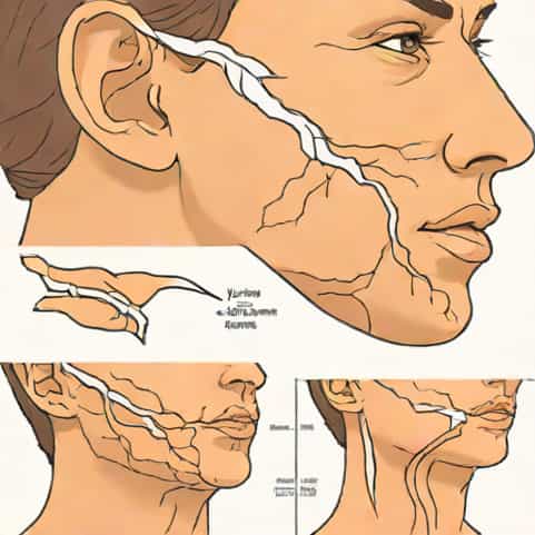
When people search for pictures of jugular vein distention, they are often trying to understand what this physical sign looks like in real life. Jugular vein distention, or JVD, is a visible bulging of the jugular veins in the neck that usually indicates increased pressure in the venous system. Doctors, nurses, medical students, and even patients may look for these images to identify the condition, recognize its causes, and understand when it signals something serious. While pictures of jugular vein distention can be helpful for learning, it is also important to understand the meaning behind what is seen, since this clinical sign often connects to heart or lung health.
What Is Jugular Vein Distention?
Jugular vein distention refers to the enlargement or visible bulging of the jugular veins, which are the veins located in the neck that carry blood back to the heart. Under normal conditions, these veins should not be overly prominent when a person is sitting upright. However, when there is increased pressure in the right atrium of the heart or in the superior vena cava, the veins may become visibly distended. Pictures of jugular vein distention often highlight this abnormal bulging along the side of the neck, making it a valuable teaching tool for medical assessment.
Why It Matters
Understanding JVD is critical because it can be a warning sign of serious health issues. When medical professionals examine patients, they often look for jugular vein distention as part of a physical exam. Photographs and diagrams help illustrate how this condition appears and how to distinguish it from other neck structures.
Causes of Jugular Vein Distention
Pictures of jugular vein distention may show similar physical appearances, but the underlying causes can vary. The most common causes include heart and lung conditions that interfere with normal blood flow. These causes explain why doctors pay close attention when they see JVD in a patient.
- Heart FailureWhen the heart is unable to pump blood effectively, pressure builds up in the veins, causing distention in the jugular veins.
- Pulmonary HypertensionHigh blood pressure in the lungs can lead to increased pressure in the right side of the heart, making jugular veins bulge.
- Cardiac TamponadeA condition where fluid builds around the heart, compressing it and leading to visible jugular vein distention.
- Tricuspid Valve DiseaseMalfunction of the tricuspid valve prevents proper blood flow, leading to backup in the jugular veins.
- Superior Vena Cava SyndromeObstruction of the major vein that carries blood to the heart can cause visible swelling in the jugular area.
Pictures of Jugular Vein Distention in Medical Education
For medical students and healthcare providers, pictures of jugular vein distention serve as essential learning materials. They demonstrate how to identify JVD during a physical exam and differentiate it from other causes of neck swelling. These images typically show patients sitting at a 45-degree angle, which is the standard position for assessing jugular venous pressure. By studying such photographs, trainees can better recognize JVD in real patients.
How JVD Appears in Pictures
In medical images, jugular vein distention usually appears as a rope-like bulge on one or both sides of the neck. It becomes most visible when the patient is reclining slightly rather than lying flat. Sometimes the distention is subtle, while in severe cases it may look like significant swelling. The clarity of these signs in pictures helps learners understand what to look for in a real clinical setting.
Clinical Examination and Pictures
Pictures of jugular vein distention also demonstrate proper examination techniques. Clinicians often compare the height of the jugular venous column with the sternal angle to estimate central venous pressure. Photographs showing this method give a clear representation of how such assessments are performed.
Steps in Recognizing JVD
While pictures give a static representation, combining them with proper technique is key
- Position the patient at a 30-45 degree angle.
- Turn the patient’s head slightly to the opposite side.
- Observe the right side of the neck for venous pulsations.
- Distinguish between jugular vein pulsations and carotid artery pulsations.
- Estimate the height of distention in relation to the sternal angle.
Limitations of Pictures
While pictures of jugular vein distention are useful, they have limitations. Lighting, patient positioning, and camera angles can affect how clearly the veins appear. In real life, motion and subtle changes in the neck can make JVD easier or harder to identify. Therefore, while photographs are excellent teaching aids, clinical practice and hands-on experience remain essential.
Why Context Is Important
A single picture of jugular vein distention does not provide the whole story. Context-such as the patient’s symptoms, medical history, and accompanying signs-is needed to interpret the meaning. For example, JVD combined with shortness of breath may suggest heart failure, while JVD with chest trauma may point toward cardiac tamponade.
When to Be Concerned
For patients, noticing bulging veins in the neck can be alarming. While pictures of jugular vein distention may help in self-recognition, it is always important to consult a healthcare professional if this sign is observed. Persistent or severe JVD often indicates a serious underlying condition that requires medical evaluation.
Other Symptoms to Watch For
When looking at pictures of jugular vein distention, people should also be aware of associated warning signs. These may include
- Shortness of breath
- Chest pain or discomfort
- Swelling in the legs or abdomen
- Rapid heartbeat or irregular pulse
- Fatigue and weakness
Educational Value of Medical Images
The value of pictures of jugular vein distention goes beyond recognition. They help build awareness of cardiovascular and pulmonary health. For medical professionals, they reinforce physical examination skills. For patients, they highlight the importance of noticing bodily changes and seeking timely medical advice.
Use in Training and Resources
Medical schools and clinical training programs often use such pictures in textbooks, lectures, and online resources. They help standardize the way clinicians identify and document JVD, improving diagnostic accuracy across different healthcare settings.
The Role of Pictures in Understanding JVD
Pictures of jugular vein distention provide a powerful way to visualize an important clinical sign. They serve as a bridge between theory and practice, allowing both students and patients to understand what JVD looks like and why it matters. Although images can be extremely helpful, they should always be interpreted with clinical context. Jugular vein distention is not just a visible change in the neck; it is a signal that something may be affecting the heart or lungs. By combining visual resources with professional evaluation, people can better understand this condition and take steps toward protecting their health.
Ultimately, while pictures of jugular vein distention help with recognition, the true value lies in what they represent an early warning sign that should never be ignored. Learning to recognize this sign can make a difference in timely diagnosis and effective treatment of serious medical conditions.