Low grade serous carcinoma is a rare but important type of ovarian cancer that often raises questions among patients, caregivers, and medical students searching for clear explanations. When people look for low grade serous carcinoma pathology outlines, they are usually trying to understand how this disease is identified under the microscope, how it differs from other ovarian tumors, and why its behavior is considered distinct. Although the term sounds highly technical, the core concepts can be explained in a way that is approachable without losing accuracy.
What Is Low Grade Serous Carcinoma
Low grade serous carcinoma is a type of epithelial ovarian cancer characterized by slow growth and relatively mild cellular abnormalities. Unlike high grade serous carcinoma, which is aggressive and fast-growing, this tumor tends to develop over a longer period of time.
It often affects younger women compared to other ovarian cancers, and its clinical course can be prolonged, sometimes lasting many years.
Why Pathology Is Central to Diagnosis
Pathology plays a critical role in diagnosing low grade serous carcinoma. Imaging studies and symptoms may suggest a mass, but the definitive diagnosis depends on microscopic examination of tissue samples.
Pathologists evaluate cell structure, growth patterns, and specific molecular features to classify the tumor accurately.
Histologic Features in Pathology Outlines
Cellular Appearance
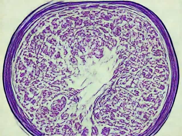
One of the defining features in low grade serous carcinoma pathology outlines is the relatively uniform appearance of tumor cells. The nuclei are small, with mild atypia, meaning they look only slightly abnormal.
This subtle appearance is a key reason why the tumor is labeled low grade.
Growth Patterns
Under the microscope, low grade serous carcinoma typically shows papillary or micropapillary structures. These finger-like projections are lined by tumor cells in an orderly fashion.
Solid sheets of cells are uncommon, which helps differentiate it from more aggressive tumors.
Mitotic Activity and Its Significance
Mitotic figures represent cell division. In low grade serous carcinoma, mitotic activity is low, meaning the cells are not dividing rapidly.
This feature supports the slow-growing nature of the disease and is a major criterion used in pathology outlines.
Psammoma Bodies
Psammoma bodies are round, calcified structures often seen in serous tumors. They are commonly present in low grade serous carcinoma.
While not exclusive to this cancer type, their presence adds supportive evidence during microscopic evaluation.
Relationship to Serous Borderline Tumors
Low grade serous carcinoma is closely related to serous borderline tumors. Many experts believe that it can develop from these borderline lesions over time.
This progression explains why the pathology outlines of both conditions share some overlapping features.
Immunohistochemistry in Diagnosis
Common Markers
Immunohistochemistry helps confirm the diagnosis by identifying specific proteins expressed by tumor cells. Low grade serous carcinoma often shows positivity for markers associated with serous differentiation.
These tests support what is seen under routine microscopy.
Hormone Receptor Expression
Many low grade serous carcinomas express estrogen and progesterone receptors. This finding has implications not only for diagnosis but also for treatment planning.
Hormone receptor positivity is one reason why hormonal therapies may be considered.
Molecular Features Highlighted in Pathology Outlines
Molecular studies have shown that low grade serous carcinoma often harbors mutations in specific signaling pathways. These mutations differ from those seen in high grade serous carcinoma.
Understanding these differences has helped refine classification and opened doors for targeted therapies.
How It Differs from High Grade Serous Carcinoma
A major goal of pathology outlines is to distinguish low grade from high grade serous carcinoma. High grade tumors show marked nuclear atypia, high mitotic rates, and chaotic growth patterns.
In contrast, low grade serous carcinoma maintains architectural order and cellular uniformity.
Clinical Correlation
Pathology findings must always be interpreted in clinical context. Low grade serous carcinoma often presents at an advanced stage, despite its slow growth.
This paradox highlights why accurate pathology assessment is essential for proper management.
Challenges in Pathologic Diagnosis
Diagnosing low grade serous carcinoma can be challenging, especially in small biopsy samples. The subtle differences between borderline tumors and invasive carcinoma require experience.
Pathologists rely on a combination of architectural patterns and evidence of stromal invasion.
Stromal Invasion
Stromal invasion is a key criterion that separates carcinoma from borderline tumors. In low grade serous carcinoma, invasion may be minimal and difficult to detect.
Careful examination of tissue sections is therefore essential.
Role of Pathology Outlines in Education
Pathology outlines serve as structured references for students and professionals. They summarize essential diagnostic criteria, differential diagnoses, and key features in a clear format.
For low grade serous carcinoma, these outlines help standardize diagnosis across institutions.
Differential Diagnosis
Several other ovarian tumors may mimic low grade serous carcinoma. These include serous borderline tumors and other low-grade epithelial neoplasms.
Pathology outlines emphasize careful evaluation to avoid misclassification.
Prognostic Implications
The pathology of low grade serous carcinoma has direct implications for prognosis. While the disease is generally indolent, it can be persistent and resistant to conventional chemotherapy.
Accurate classification helps guide long-term management strategies.
Treatment Considerations Linked to Pathology
Because of its unique biology, low grade serous carcinoma may respond differently to treatments compared to high grade tumors.
Pathology findings influence decisions regarding surgery, hormonal therapy, and targeted treatments.
Ongoing Research and Evolving Criteria
Research continues to refine the pathology outlines of low grade serous carcinoma. New molecular markers and classification systems are being studied.
These advances aim to improve diagnostic accuracy and patient outcomes.
Low grade serous carcinoma pathology outlines provide a structured way to understand this distinct ovarian cancer. By focusing on cellular uniformity, low mitotic activity, specific growth patterns, and molecular features, pathologists can make accurate diagnoses.
For readers seeking clarity, understanding these pathology principles helps demystify the disease and highlights why precise classification is essential in modern cancer care.