The female Anopheles mosquito is a significant insect in medical entomology because of its role as the primary vector for malaria. Understanding its anatomy is crucial for students, researchers, and health professionals who study mosquito behavior, disease transmission, and control measures. A labelled diagram of the female Anopheles mosquito helps in identifying key parts of its body, understanding their functions, and appreciating how its structure enables it to transmit malaria. By examining the mosquito’s head, thorax, abdomen, wings, and legs, we can gain insight into how it feeds, reproduces, and interacts with its environment.
Introduction to Female Anopheles Mosquito
Female Anopheles mosquitoes differ from males primarily in their ability to bite and transmit disease. Only females require blood meals to develop eggs, while males feed on nectar. The female mosquito’s anatomy is adapted for blood-feeding and survival, making it an efficient vector for malaria. Studying a labelled diagram allows students to recognize features like the proboscis, antennae, and other critical structures that contribute to its role in disease transmission. Accurate identification of these features is also essential for implementing targeted mosquito control strategies.
Importance in Malaria Transmission
The female Anopheles mosquito is medically important because it transmits the Plasmodium parasite, which causes malaria. The anatomical features, such as the elongated proboscis and specialized salivary glands, are directly related to its ability to extract blood from hosts and inject the parasite. A clear understanding of these anatomical structures helps researchers develop interventions like insecticide-treated nets, repellents, and other control measures.
Key Features in the Labelled Diagram
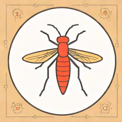
A labelled diagram of the female Anopheles mosquito provides a visual representation of its major anatomical parts. Each part has a specific function that contributes to the mosquito’s survival, reproduction, and role as a disease vector.
Head
The head of the female Anopheles mosquito contains sensory organs and feeding structures.
- ProboscisAn elongated, needle-like structure used to pierce the skin and suck blood. It contains two tubes; one for injecting saliva and the other for drawing blood.
- AntennaeLong, segmented organs that detect carbon dioxide, body heat, and odors, helping the mosquito locate hosts.
- Compound EyesLarge, multifaceted eyes that provide a wide field of vision and detect movement, which aids in host detection and navigation.
- PalpsSensory appendages that help the mosquito detect temperature and chemical cues from potential hosts.
Thorax
The thorax is the central body segment that houses muscles essential for flight and locomotion.
- WingsTwo transparent wings that enable flight. The wings’ movement allows the mosquito to reach hosts and escape predators.
- LegsSix long, segmented legs attached to the thorax. The legs are used for landing, walking on surfaces, and sensing vibrations.
- Flight MusclesLocated inside the thorax, these powerful muscles control wing movement and enable sustained flight necessary for host-seeking and oviposition.
Abdomen
The abdomen is the posterior segment of the mosquito and is primarily involved in digestion, reproduction, and blood storage.
- Digestive SystemContains the midgut where blood is digested and nutrients are absorbed. The midgut is also where the malaria parasite develops.
- OvariesFemale reproductive organs that produce eggs after blood meals. The ovaries are crucial for the continuation of the mosquito population.
- Salivary GlandsSecrete anticoagulants and enzymes to facilitate blood-feeding and are the site where Plasmodium sporozoites are stored before transmission to a host.
- SpiraclesSmall openings on the abdomen used for respiration.
Functions of Major Anatomical Parts
Each labeled structure in the female Anopheles mosquito plays a vital role in its survival and its ability to transmit malaria.
Proboscis and Feeding
The proboscis allows the female mosquito to extract blood efficiently. Its dual-tube system ensures that saliva, which contains anticoagulants and potentially malaria parasites, is injected while blood is ingested. Understanding the proboscis’s structure is essential for designing repellents and other preventive measures.
Antennae and Sensory Organs
Antennae and palps detect chemical and thermal signals emitted by hosts. This sensitivity helps mosquitoes locate humans and animals, ensuring successful blood-feeding for egg development.
Flight and Mobility
The thorax houses flight muscles and supports wings and legs, enabling movement and host-seeking. Mosquitoes rely on these structures to find suitable feeding sites and avoid threats, which makes them highly adaptable and efficient vectors.
Reproduction and Egg Development
The abdomen contains the ovaries and other reproductive structures. After consuming a blood meal, the female mosquito develops eggs and lays them in water bodies. Understanding this process is critical for controlling mosquito populations and preventing malaria transmission.
Importance of Labelled Diagrams in Education
Labelled diagrams of the female Anopheles mosquito are invaluable educational tools for students, researchers, and public health professionals. They provide a clear visual representation of complex anatomical structures and help learners understand how the mosquito functions as a vector for disease.
Benefits for Students
- Facilitates easier memorization of mosquito anatomy
- Illustrates the relationship between structure and function
- Supports practical learning for entomology and biology studies
Benefits for Researchers and Health Professionals
- Helps in identifying target points for vector control
- Assists in understanding parasite transmission mechanisms
- Guides the development of insecticides, repellents, and other interventions
Applications in Malaria Control
Understanding the anatomy of the female Anopheles mosquito through labelled diagrams contributes directly to malaria prevention strategies. By identifying key anatomical features, scientists can develop targeted interventions that disrupt blood-feeding, reproduction, or parasite development.
Vector Control Strategies
Labelled diagrams aid in the design of tools such as
- Insecticide-treated nets (ITNs) that prevent mosquitoes from reaching hosts
- Repellents that target sensory organs like antennae and palps
- Larvicides and environmental interventions targeting breeding sites in water bodies
Research and Monitoring
Researchers use knowledge of mosquito anatomy to monitor populations, study behavior, and assess the effectiveness of control measures. Understanding the female mosquito’s anatomy is essential since it is the primary disease vector, unlike males which do not feed on blood.
A labelled diagram of the female Anopheles mosquito provides an essential tool for understanding its anatomy, behavior, and role as a vector for malaria. The head, thorax, and abdomen contain specialized structures that enable blood-feeding, reproduction, and flight, making the mosquito an efficient carrier of the Plasmodium parasite. Educationally, these diagrams help students and health professionals visualize complex anatomical details, while practically, they support research, vector control, and public health interventions. By studying the labelled diagram, we gain insight into the biology of one of the world’s most important disease vectors and can develop strategies to reduce the global burden of malaria.