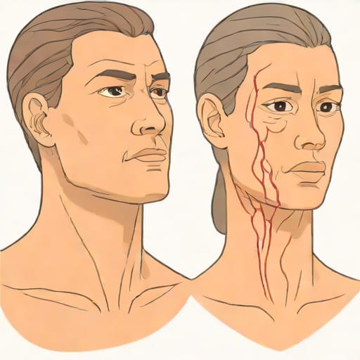
Jugular vein distention (JVD) is a physical sign that healthcare professionals often examine when assessing a patient’s cardiovascular health. It refers to the abnormal swelling or bulging of the jugular vein in the neck, which can indicate elevated central venous pressure. In contrast, a normal jugular vein is usually flat or only slightly visible when a person is sitting or lying down at a 45-degree angle. Understanding the differences between jugular vein distention and normal jugular vein presentation is crucial for early diagnosis of heart conditions such as congestive heart failure, pulmonary hypertension, or fluid overload. By carefully observing and interpreting these signs, clinicians can make informed decisions about patient care and treatment options.
Understanding Jugular Vein Anatomy
Location and Function
The jugular veins are major blood vessels that carry deoxygenated blood from the head and neck back to the heart. There are two main types the internal jugular vein and the external jugular vein. The internal jugular vein runs alongside the carotid artery and drains directly into the superior vena cava, while the external jugular vein is more superficial and drains the outer portions of the head and neck. The primary function of these veins is to regulate blood flow toward the heart, making them key indicators of central venous pressure and overall cardiovascular health.
Normal Jugular Vein Appearance
In a healthy individual, the jugular vein is not prominently visible when the person is in a sitting position. The internal jugular vein may only be slightly noticeable when lying down with the head elevated at a 30 to 45-degree angle. A normal jugular vein exhibits pulsations synchronized with the heartbeat, typically reflecting right atrial pressure. These subtle pulsations are usually easy for trained clinicians to identify and serve as a baseline for comparison when evaluating potential distention.
What Is Jugular Vein Distention?
Definition and Clinical Significance
Jugular vein distention occurs when the jugular vein becomes visibly enlarged or prominent due to increased pressure in the right atrium and superior vena cava. This condition often signals that the heart is struggling to manage blood flow, which can be a hallmark of underlying cardiovascular problems. Clinicians view JVD as a non-invasive method to assess the severity of heart failure or other circulatory issues, making it an important diagnostic tool in both emergency and routine clinical settings.
Causes of Jugular Vein Distention
Several conditions can lead to jugular vein distention, including
- Congestive heart failure where the heart cannot pump blood effectively, causing fluid buildup.
- Pulmonary hypertension increased pressure in the pulmonary arteries can affect venous return.
- Tricuspid valve disease malfunctioning heart valves can lead to backflow and vein distention.
- Pericardial tamponade accumulation of fluid around the heart restricts its pumping ability.
- Chronic kidney disease fluid overload may contribute to elevated central venous pressure.
Recognizing these potential causes allows healthcare providers to initiate further diagnostic testing and interventions promptly, improving patient outcomes.
How to Assess Jugular Vein Distention
Patient Positioning
Proper assessment of jugular vein distention begins with correct patient positioning. The patient should be lying on their back with the upper body elevated at a 30 to 45-degree angle. The head should be turned slightly to the opposite side of the vein being examined. Good lighting is essential to visualize the vein clearly. By positioning the patient correctly, clinicians can distinguish between normal jugular pulsations and abnormal distention.
Measurement Techniques
Clinicians often measure the vertical height of the jugular vein from the sternal angle to the top of the venous column. A measurement greater than 3 centimeters above the sternal angle is usually considered indicative of jugular vein distention. Observing the waveform of the vein is also important, as abnormal pulsations can provide clues about underlying heart conditions. Combining visual inspection with palpation helps ensure accurate assessment.
Differences Between Jugular Vein Distention and Normal Vein
Visual Appearance
A normal jugular vein is subtle and may only be visible under specific conditions, showing gentle pulsations synchronized with the heartbeat. In contrast, jugular vein distention is characterized by visible bulging that persists even when the patient is sitting upright. The vein may appear swollen and more prominent on one or both sides of the neck, signaling increased central venous pressure.
Clinical Implications
While a normal jugular vein indicates healthy central venous pressure and cardiac function, distention is often a warning sign of cardiovascular stress. Persistent JVD can indicate acute or chronic heart problems, fluid overload, or other systemic issues requiring immediate attention. Recognizing these differences allows healthcare providers to differentiate between benign variations and conditions that necessitate urgent care.
Associated Symptoms and Related Findings
Common Symptoms
Patients with jugular vein distention may also present with additional symptoms, including
- Shortness of breath or difficulty breathing
- Swelling in the legs or ankles
- Fatigue or weakness
- Rapid or irregular heartbeat
- Chest pain or pressure in severe cases
Recognizing these accompanying symptoms helps clinicians assess the severity and urgency of the condition.
Diagnostic Testing
To confirm the cause of jugular vein distention, healthcare providers may order diagnostic tests such as echocardiography, chest X-rays, electrocardiograms (ECG), or blood tests. These tests help identify underlying heart dysfunction, fluid overload, or other systemic conditions contributing to elevated venous pressure. Proper diagnosis is essential for effective treatment planning.
Treatment and Management
Addressing the Underlying Cause
Treatment for jugular vein distention focuses on resolving the underlying condition. For example, managing congestive heart failure may involve diuretics, ACE inhibitors, or lifestyle modifications such as reduced salt intake. In cases of valvular heart disease, surgical intervention may be required. Timely and appropriate treatment can reduce JVD and improve overall cardiovascular health.
Monitoring and Follow-Up
Regular monitoring of jugular vein appearance and central venous pressure is important for patients with known heart conditions. Follow-up appointments allow clinicians to track progress, adjust medications, and intervene early if symptoms worsen. Patient education on recognizing changes in vein distention and associated symptoms also plays a crucial role in long-term management.
Understanding the differences between jugular vein distention and a normal jugular vein is essential for accurate cardiovascular assessment. While a normal jugular vein reflects healthy venous pressure and cardiac function, JVD can indicate serious underlying conditions such as heart failure, pulmonary hypertension, or fluid overload. Careful examination, patient positioning, and measurement techniques allow clinicians to identify distention and initiate appropriate diagnostic and therapeutic measures. Recognizing these signs, alongside associated symptoms and diagnostic findings, is critical for effective patient care, early intervention, and improved cardiovascular outcomes.