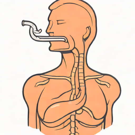
Thickening of the esophagus is a term often encountered during medical imaging or endoscopic examinations, and it naturally raises concerns about the possibility of cancer. The esophagus is a muscular tube that connects the throat to the stomach, and changes in its wall thickness can result from a variety of conditions, both benign and malignant. While cancer is one potential cause, it is not the only explanation for esophageal thickening. Understanding what thickening of the esophagus means requires a careful review of symptoms, imaging findings, medical history, and additional diagnostic tests. Early detection and evaluation are critical for determining the cause and initiating appropriate treatment.
Understanding Esophageal Thickening
Esophageal thickening refers to an increase in the thickness of the esophageal wall observed on imaging studies such as a CT scan, MRI, or endoscopic ultrasound. The esophagus normally has a thin, smooth wall that allows food to pass efficiently from the mouth to the stomach. When the wall becomes thicker than usual, it may indicate inflammation, injury, or abnormal growth of tissue. The location, symmetry, and extent of thickening are important factors in evaluating the cause.
Possible Causes of Esophageal Thickening
Not all cases of esophageal thickening indicate cancer. There are several benign conditions that can lead to thickening of the esophagus
- Gastroesophageal reflux disease (GERD)Chronic acid exposure can cause inflammation, leading to thickened esophageal walls.
- EsophagitisInflammation due to infection, irritation, or autoimmune disorders may cause localized or diffuse thickening.
- Esophageal stricturesScar tissue from chronic irritation or previous injury can thicken the esophageal wall.
- Motility disordersConditions such as achalasia can cause muscular thickening of the esophagus due to chronic dysfunction.
- Benign tumorsNoncancerous growths like leiomyomas can appear as focal thickening on imaging.
Esophageal Cancer and Wall Thickening
While thickening of the esophagus can be caused by benign conditions, it can also be a sign of esophageal cancer. Esophageal cancer typically presents as a localized area of irregular, asymmetric thickening rather than uniform thickening along the entire length of the esophagus. The two main types of esophageal cancer are squamous cell carcinoma and adenocarcinoma, each with different risk factors and locations within the esophagus.
Risk Factors for Esophageal Cancer
- Chronic acid reflux and Barrett’s esophagus
- Smoking and heavy alcohol consumption
- Obesity and dietary factors
- History of esophageal injury or radiation exposure
- Genetic predisposition in some families
Symptoms Suggestive of Cancer
Certain symptoms accompanying esophageal thickening may raise suspicion for malignancy
- Difficulty swallowing (dysphagia), especially for solids initially
- Unexplained weight loss
- Persistent chest pain or discomfort
- Vomiting or regurgitation of food
- Chronic hoarseness or cough in advanced cases
Diagnostic Evaluation
Determining whether esophageal thickening represents cancer requires a systematic evaluation. Physicians often start with imaging studies and endoscopic assessments to visualize the esophagus and take tissue samples if necessary.
Imaging Studies
CT scans, MRI, and barium swallow studies can identify areas of thickening and help differentiate between diffuse and focal changes. Irregular, asymmetric thickening with mass formation is more concerning for cancer, whereas uniform, smooth thickening is often benign.
Endoscopy and Biopsy
Endoscopy allows direct visualization of the esophageal lining and enables biopsy of suspicious areas. A biopsy is essential for a definitive diagnosis, as imaging alone cannot distinguish cancer from inflammation or benign growths.
Endoscopic Ultrasound (EUS)
Endoscopic ultrasound provides detailed information about the layers of the esophageal wall. This technique can assess how deep a lesion extends and whether nearby lymph nodes are involved, which is crucial for staging esophageal cancer if present.
Management Based on Cause
Treatment depends entirely on the underlying cause of esophageal thickening. For benign conditions such as esophagitis or reflux-induced changes, management focuses on reducing inflammation and preventing further injury. Lifestyle modifications, medications like proton pump inhibitors, and dilation procedures for strictures are commonly employed. In contrast, malignant thickening requires oncologic evaluation and treatment, which may include surgery, chemotherapy, radiation therapy, or a combination of these approaches.
Monitoring and Follow-Up
Even if initial evaluation suggests a benign cause, follow-up may be necessary. Chronic inflammation or Barrett’s esophagus, for example, can increase the risk of developing cancer over time. Regular endoscopic surveillance allows early detection and intervention for precancerous changes.
Key Takeaways
Thickening of the esophagus does not automatically mean cancer. It can result from a variety of conditions, including inflammation, scarring, motility disorders, benign tumors, and malignant growths. The characteristics of the thickening, associated symptoms, and risk factors help guide the evaluation. Definitive diagnosis requires imaging studies, endoscopy, and biopsy. Timely assessment and monitoring are essential to ensure that serious conditions are detected early while benign causes are managed appropriately.
- Esophageal thickening can be benign or malignant.
- Imaging studies help identify the pattern and extent of thickening.
- Endoscopy with biopsy is required for a definitive diagnosis.
- Symptoms, risk factors, and patient history guide clinical suspicion.
- Management depends on the underlying cause, ranging from lifestyle changes to cancer treatment.
In summary, while thickening of the esophagus can be a sign of cancer, it is not definitive on its own. Many noncancerous conditions can lead to similar findings on imaging. Proper evaluation with a combination of imaging, endoscopy, and biopsy is essential to determine the cause. Understanding the difference between benign and malignant causes allows for appropriate management and timely intervention, reducing the risk of complications and improving outcomes. Anyone experiencing symptoms such as difficulty swallowing, unexplained weight loss, or persistent chest discomfort should seek medical attention promptly for proper assessment and care.