The knee is one of the most complex and important joints in the human body, responsible for supporting body weight, enabling movement, and maintaining stability. Among its critical components are the collateral ligaments, which are essential for controlling side-to-side motion and preventing excessive lateral or medial displacement. Understanding where a collateral ligament of the knee is located, its function, and the injuries associated with it is crucial for medical students, athletes, and anyone interested in musculoskeletal health. These ligaments, along with other stabilizing structures such as the anterior and posterior cruciate ligaments, menisci, and muscles, ensure proper knee mechanics during activities like walking, running, and jumping.
Definition and Types of Collateral Ligaments
Collateral ligaments are strong bands of connective tissue that connect bones and provide stability to the knee joint. There are two main collateral ligaments in the knee
- Medial Collateral Ligament (MCL)Located on the inner side of the knee, the MCL connects the femur (thigh bone) to the tibia (shin bone). Its primary function is to resist forces that push the knee inward, known as valgus forces.
- Lateral Collateral Ligament (LCL)Located on the outer side of the knee, the LCL connects the femur to the fibula, a smaller bone alongside the tibia. The LCL helps prevent excessive outward bending, or varus forces, stabilizing the outer aspect of the knee.
Location of the Medial Collateral Ligament (MCL)
The MCL is positioned along the inner side of the knee and can be felt when touching the medial side of the joint. It extends from the medial epicondyle of the femur down to the medial condyle and shaft of the tibia. The MCL consists of superficial and deep fibers. The superficial fibers provide major resistance to valgus stress, while the deep fibers are connected to the medial meniscus, helping coordinate stability between the ligament and the meniscus. This location makes the MCL particularly susceptible to injury during sports that involve sudden changes in direction or direct impact to the lateral side of the knee.
Location of the Lateral Collateral Ligament (LCL)
The LCL is found along the outer side of the knee and is more slender compared to the MCL. It runs from the lateral epicondyle of the femur to the head of the fibula. Unlike the MCL, the LCL does not attach directly to the meniscus. It primarily resists varus forces that push the knee outward. Its positioning allows it to stabilize the knee against rotational forces and helps prevent excessive side-to-side motion. Injuries to the LCL are less common than MCL injuries but can occur during high-impact collisions or activities that involve twisting motions of the knee.
Functions of Collateral Ligaments
Collateral ligaments play a vital role in maintaining knee stability and overall joint function. Their location on the medial and lateral sides of the knee allows them to perform several critical functions
- Side-to-Side StabilityPreventing the knee from bending excessively inward (valgus) or outward (varus).
- Coordination with MenisciEspecially the MCL, which connects to the medial meniscus, supporting proper load distribution across the knee joint.
- Protection During MovementHelping the knee withstand forces during running, jumping, and sudden directional changes.
- Rotational StabilityAssisting other ligaments and muscles in controlling rotational movements and preventing torsional injuries.
Common Injuries to Collateral Ligaments
Collateral ligament injuries are frequent in sports and everyday activities, especially in high-contact sports like football, soccer, and skiing. MCL injuries are more common than LCL injuries due to their anatomical location and exposure to medial stress. Injuries can range from mild sprains to complete tears and are usually classified in grades
- Grade 1Mild stretching without significant tearing, minimal instability.
- Grade 2Partial tear with moderate pain, swelling, and slight instability.
- Grade 3Complete tear resulting in significant instability and difficulty bearing weight.
Causes of MCL and LCL Injuries
- Direct blow to the lateral side of the knee (MCL injuries).
- Direct blow to the medial side of the knee (LCL injuries).
- Sudden twisting or pivoting movements, often seen in soccer or basketball.
- Falls or collisions in skiing or high-impact sports.
Symptoms of Collateral Ligament Injuries
- Pain along the inner or outer knee depending on the ligament injured.
- Swelling and tenderness along the ligament line.
- Instability or a feeling that the knee gives out.
- Limited range of motion in severe injuries.
Diagnosis and Imaging
Identifying the location and severity of a collateral ligament injury requires a combination of physical examination and imaging studies. Physicians often perform valgus and varus stress tests to assess the MCL and LCL, respectively. Swelling and tenderness along the ligament help localize the injury. Advanced imaging such as MRI is commonly used to visualize the ligament’s fibers, detect partial or complete tears, and evaluate associated injuries to menisci or other ligaments. Accurate diagnosis is essential to determine the appropriate treatment plan and prevent long-term knee instability.
Physical Examination Techniques
- Valgus Stress TestEvaluates MCL stability by applying inward pressure on the lateral knee.
- Varus Stress TestEvaluates LCL stability by applying outward pressure on the medial knee.
- PalpationLocating tenderness along the medial or lateral sides to pinpoint ligament involvement.
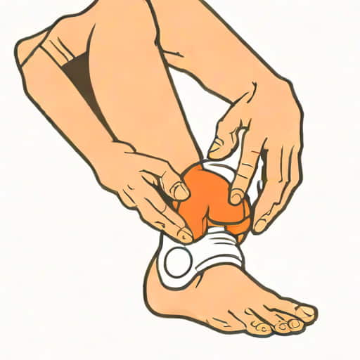
Treatment Options
Treatment depends on the location, severity, and overall condition of the knee. Mild injuries often respond well to conservative treatments, while severe injuries may require surgical intervention.
Conservative Management
- Rest and activity modification to allow the ligament to heal.
- Ice therapy to reduce swelling and pain.
- Compression and elevation to minimize inflammation.
- Physical therapy to restore strength, stability, and range of motion.
Surgical Intervention
Severe grade 3 tears, chronic instability, or combined injuries with other ligaments may require reconstructive surgery. Surgical repair often involves stitching torn fibers or grafting tissue from other tendons to restore stability. Postoperative rehabilitation focuses on gradually regaining mobility and strengthening the surrounding muscles to support the ligament.
Prevention and Care
Preventing collateral ligament injuries involves strengthening the muscles around the knee, improving flexibility, and using proper techniques during sports and physical activities. Wearing protective gear, avoiding sudden twists or excessive lateral forces, and maintaining overall fitness can significantly reduce the risk of injury. Regular warm-up exercises, balance training, and controlled stretching are essential for athletes and active individuals to maintain ligament health and knee stability.
A collateral ligament of the knee, whether the medial collateral ligament (MCL) or lateral collateral ligament (LCL), is located on the inner and outer sides of the knee, respectively. These ligaments provide essential stability, preventing excessive side-to-side movements and contributing to rotational control. Understanding their location, function, and potential injuries is crucial for anyone involved in sports, physical therapy, or medical studies. Injuries can range from mild sprains to complete tears, with symptoms including pain, swelling, and instability. Accurate diagnosis through physical exams and imaging helps guide treatment, which may involve conservative care or surgical repair. Preventive measures, such as strengthening exercises and proper sports techniques, play a vital role in protecting these ligaments. By knowing where a collateral ligament of the knee is located and its role in joint stability, individuals can better appreciate the complexity of knee mechanics and take steps to maintain optimal knee health.