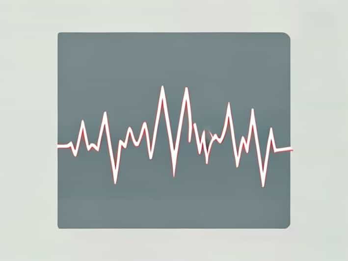
An electrocardiogram, commonly called an EKG or ECG, is a test that records the electrical activity of the heart. It is a crucial tool in diagnosing heart conditions, as it provides insight into the rhythm, rate, and overall health of the heart. Understanding how an abnormal EKG looks can help patients and healthcare providers recognize potential problems early. Abnormalities in an EKG can indicate issues ranging from irregular heart rhythms to heart attacks, electrolyte imbalances, or structural heart disease, making this knowledge essential for proper evaluation and treatment.
Basics of a Normal EKG
Before exploring abnormal EKGs, it is important to understand what a normal EKG looks like. A standard EKG tracing consists of several components
- P waveRepresents atrial depolarization, the electrical activity that causes the atria to contract.
- QRS complexIndicates ventricular depolarization, which triggers the main pumping chambers to contract.
- T waveRepresents ventricular repolarization, the recovery phase of the ventricles.
- PR intervalThe time from the start of the P wave to the start of the QRS complex, showing how long it takes for the electrical signal to travel from the atria to the ventricles.
- ST segmentThe flat section between the QRS complex and the T wave, often analyzed for signs of ischemia or heart attack.
In a normal EKG, these waves are consistent, with regular spacing and duration, reflecting a healthy and coordinated heart rhythm.
Common Abnormal EKG Patterns
An abnormal EKG may appear in various ways depending on the underlying heart condition. Some abnormalities are subtle, while others are obvious, making interpretation a skill that requires clinical expertise. Here are some of the most common patterns seen in abnormal EKGs.
Arrhythmias (Irregular Heart Rhythms)
Arrhythmias are one of the most frequent causes of abnormal EKGs. They occur when the heart beats too fast, too slow, or irregularly.
- Atrial fibrillationCharacterized by a lack of distinct P waves and an irregularly irregular rhythm. The heart rate can be fast or slow, and the rhythm is chaotic.
- Ventricular tachycardiaShows a series of wide, rapid QRS complexes without preceding P waves. This is a serious condition that may require immediate treatment.
- BradycardiaA slow heart rate, often with regular intervals but abnormally spaced QRS complexes.
Signs of Myocardial Infarction (Heart Attack)
Abnormal EKGs can indicate a heart attack by showing characteristic changes in the ST segment, T waves, or Q waves.
- ST elevationThe ST segment rises above the baseline, often in specific leads, signaling an acute heart attack.
- ST depressionThe ST segment falls below the baseline, indicating ischemia or insufficient blood flow to the heart.
- Pathological Q wavesDeep and wide Q waves may appear after a previous heart attack, representing areas of dead heart tissue.
Conduction Abnormalities
Problems with the heart’s electrical conduction system can also cause abnormal EKGs. These include
- Bundle branch blocksWide QRS complexes with characteristic patterns, indicating delayed electrical conduction through the ventricles.
- Heart blocksAbnormal PR intervals suggest issues with atrioventricular conduction. First-degree blocks have prolonged PR intervals, while second- and third-degree blocks show intermittent or complete failure of conduction.
Electrolyte Imbalances and Medication Effects
Certain abnormalities in an EKG may reflect changes in potassium, calcium, or magnesium levels, or the effects of medications like digoxin or antiarrhythmics.
- HyperkalemiaPeaked T waves, flattened P waves, and widened QRS complexes.
- HypokalemiaFlattened T waves, prominent U waves, and ST depression.
- Drug-induced changesSome medications prolong the QT interval, increasing the risk of dangerous arrhythmias.
ST Segment and T Wave Abnormalities
The ST segment and T wave are crucial for detecting ischemia and other heart conditions. Abnormalities here can indicate
- ST elevation or depression, signaling myocardial injury or ischemia.
- Inverted T waves, often associated with ischemia or previous heart damage.
- Flattened or biphasic T waves, which may suggest electrolyte imbalances or conduction problems.
QT Interval Abnormalities
The QT interval, measured from the start of the QRS complex to the end of the T wave, reflects the time the ventricles take to depolarize and repolarize. Abnormalities include
- Prolonged QTCan lead to torsades de pointes, a potentially life-threatening arrhythmia.
- Shortened QTMay indicate conditions like hypercalcemia or congenital short QT syndrome.
Recognizing Abnormal EKGs in Practice
Healthcare providers look at the overall pattern, spacing, and morphology of the waves to identify abnormalities. While some patterns suggest minor issues, others require immediate intervention. For instance, ST elevation or ventricular tachycardia are considered emergencies, whereas slight PR interval prolongation might be monitored over time.
It is also important to compare with previous EKGs when available, as some variations may be normal for certain individuals or related to age, athletic conditioning, or medications.
Red Flags on an EKG
- Irregular or absent P waves
- Wide or bizarre QRS complexes
- ST segment elevation or depression
- Abnormally long or short QT interval
- Prominent U waves or inverted T waves
Understanding how an abnormal EKG looks is essential for early detection and management of heart conditions. Abnormalities can indicate arrhythmias, myocardial infarction, conduction problems, electrolyte imbalances, or medication effects. Key signs include irregular rhythms, ST segment deviations, T wave changes, and QT interval abnormalities. While recognizing these patterns requires clinical training, awareness of what constitutes a normal versus abnormal EKG helps patients understand why this test is vital for cardiovascular health.
Regular monitoring, timely interpretation, and consultation with healthcare providers ensure that abnormal EKG findings are addressed appropriately. An EKG remains a powerful, noninvasive tool for detecting heart problems before they become life-threatening, making knowledge of abnormal patterns crucial for both medical professionals and informed patients.