Lumbago, commonly referred to as lower back pain, is one of the most frequent musculoskeletal complaints worldwide. It can range from mild discomfort to debilitating pain that affects daily activities and quality of life. Diagnosing the exact cause of lumbago often involves imaging studies, among which X-rays are commonly used. X-ray findings can provide valuable information about the structural integrity of the lumbar spine, revealing abnormalities such as vertebral degeneration, fractures, or alignment issues. Understanding these findings is crucial for healthcare providers to develop an effective treatment plan and for patients to comprehend the underlying factors contributing to their pain.
Understanding Lumbago
Lumbago refers to pain localized in the lower back area, which may involve muscles, ligaments, intervertebral discs, nerves, or vertebrae. It can be classified as acute, subacute, or chronic depending on the duration of symptoms. Causes of lumbago vary widely, ranging from simple muscle strain to degenerative spinal conditions or traumatic injuries. While clinical evaluation and physical examination provide essential insights, imaging studies like X-rays help confirm structural abnormalities and guide management.
Common Causes of Lumbago
- Muscle strain or ligament sprain from overuse or sudden movements.
- Degenerative changes in intervertebral discs or facet joints.
- Herniated discs compressing spinal nerves.
- Osteoporosis or vertebral compression fractures.
- Scoliosis or abnormal spinal alignment.
- Infections or tumors, though less common.
The Role of X-Rays in Lumbago
X-rays are a primary imaging modality used to evaluate the bony structures of the lumbar spine. They are widely available, cost-effective, and provide quick information about vertebral anatomy. X-rays are particularly useful for detecting fractures, degenerative changes, spinal alignment issues, and congenital abnormalities. However, they have limitations in assessing soft tissues, such as muscles, ligaments, and intervertebral discs, which may require MRI or CT scans for more detailed evaluation.
Indications for Lumbar Spine X-Rays
Healthcare providers may order X-rays in patients with lumbago under certain circumstances, including
- Persistent lower back pain lasting more than six weeks.
- History of trauma or injury to the lumbar spine.
- Neurological symptoms such as numbness, tingling, or weakness in the legs.
- Suspected fractures or spinal deformities.
- Evaluation of degenerative or osteoarthritic changes.
Typical X-Ray Findings in Lumbago
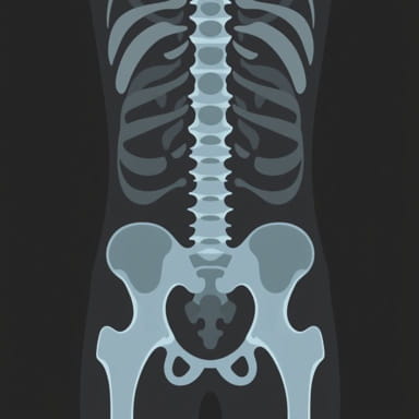
Interpreting X-ray findings requires careful assessment of vertebral bodies, intervertebral disc spaces, facet joints, and spinal alignment. Common findings include
1. Degenerative Changes
Degenerative changes in the lumbar spine are frequent in adults and may be observed as
- Narrowing of intervertebral disc spaces due to disc degeneration.
- Osteophytes or bone spurs along the vertebral margins.
- Sclerosis of the vertebral endplates.
These changes may correlate with chronic lower back pain and stiffness, although some individuals may remain asymptomatic despite radiographic findings.
2. Vertebral Fractures
X-rays can detect compression or traumatic fractures in the vertebrae. Compression fractures are common in patients with osteoporosis and may present as wedge-shaped vertebrae on lateral X-rays. Traumatic fractures from accidents or falls can also be identified, often requiring urgent medical attention.
3. Spinal Alignment Abnormalities
Spinal deformities such as scoliosis, kyphosis, or lordosis can be evaluated using X-rays. Abnormal curvature of the spine may contribute to lumbago and affect posture, gait, and overall spinal mechanics. X-rays help quantify the degree of curvature and monitor progression over time.
4. Spondylolisthesis
Spondylolisthesis occurs when one vertebra slips forward relative to the one below it. X-rays can reveal the degree of slippage and its potential impact on spinal stability. This condition may cause lower back pain, nerve compression, and limited mobility.
5. Signs of Infection or Tumor
Although rare, X-rays may detect abnormal bone destruction or lesions suggesting infection (osteomyelitis) or tumor presence. In such cases, further imaging with MRI or CT and laboratory testing is often necessary for definitive diagnosis and treatment planning.
Limitations of X-Rays in Lumbago Evaluation
While X-rays provide valuable information about bone structures, they have certain limitations
- Limited visualization of soft tissues such as discs, muscles, ligaments, and nerves.
- Early degenerative changes or minor disc herniations may not be apparent.
- Radiographic findings do not always correlate with the severity of pain.
Therefore, X-rays are often used in combination with clinical evaluation and, if necessary, advanced imaging modalities to provide a comprehensive assessment of lumbago.
Clinical Correlation and Management
X-ray findings must be interpreted in conjunction with the patient’s symptoms, medical history, and physical examination. Not all radiographic abnormalities cause pain, and some individuals with significant structural changes may remain symptom-free. Management strategies based on X-ray findings may include
Conservative Treatments
- Rest and activity modification to reduce stress on the lumbar spine.
- Physical therapy focusing on strengthening core muscles and improving flexibility.
- Pain relief with medications such as NSAIDs or muscle relaxants.
- Heat or cold therapy for symptomatic relief.
Interventional and Surgical Options
In cases where structural abnormalities are severe or conservative treatment fails, interventional procedures or surgery may be considered
- Epidural steroid injections for nerve compression or inflammation.
- Surgical decompression or spinal fusion for fractures, severe spondylolisthesis, or spinal instability.
- Minimally invasive procedures to address disc herniation or bone spurs.
Preventive Measures and Lifestyle Considerations
Preventing lumbago and minimizing progression of structural spinal changes involves lifestyle modifications and proactive care
- Maintaining a healthy weight to reduce stress on the lumbar spine.
- Regular exercise, including core strengthening and flexibility training.
- Ergonomic adjustments at work and proper lifting techniques.
- Avoiding prolonged periods of sitting or standing without movement.
- Timely management of underlying conditions such as osteoporosis or arthritis.
Lumbago X-ray findings provide critical insights into the structural integrity of the lumbar spine and help guide the diagnosis and management of lower back pain. Common findings include degenerative changes, vertebral fractures, spinal alignment abnormalities, spondylolisthesis, and occasionally signs of infection or tumor. While X-rays are a valuable diagnostic tool, they have limitations, particularly in evaluating soft tissue structures, and should be interpreted alongside clinical assessment. Management strategies range from conservative approaches like rest, physical therapy, and medications to interventional procedures or surgery for severe cases. Preventive measures, lifestyle modifications, and proactive care play an essential role in reducing the risk of recurrent lumbago and maintaining spinal health. Understanding X-ray findings in the context of symptoms empowers patients and healthcare providers to make informed decisions and achieve optimal outcomes for lower back pain.