The relationship between different structures in the human body can be understood more clearly by using anatomical directional terms. These standardized terms allow students, healthcare workers, and anyone studying anatomy to describe the precise location of one structure relative to another. One common comparison used in basic anatomy lessons is the position of the vertebral column relative to the trachea. Because these structures lie close together in the neck and upper thoracic region, understanding their directional orientation helps build a strong foundation for learning about body systems, medical examinations, and common clinical procedures.
Understanding the Vertebral Column and Trachea
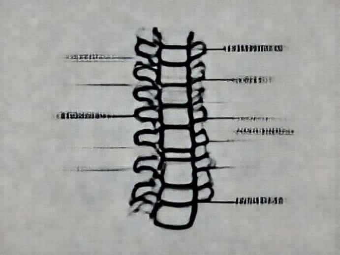
The vertebral column, often called the spine, forms the central supporting framework of the body. It protects the spinal cord, supports posture, and connects various skeletal structures. The trachea, on the other hand, is part of the respiratory system and functions as the airway that carries air from the larynx to the lungs. Although they serve very different purposes, their anatomical relationship is essential for understanding the organization of the cervical region.
General Location of Both Structures
The vertebral column runs down the midline of the back, forming the posterior part of the torso. The trachea is positioned in the anterior portion of the neck and chest. Because of this, learning the proper directional terms to describe their orientation becomes much easier once you visualize their placement relative to each other.
Key Directional Terms in Anatomy
Directional terms in anatomy help describe positions without confusion. These terms are universal, meaning they remain consistent regardless of the observer’s perspective. When comparing the vertebral column to the trachea, several primary terms become relevant.
- Anterior (ventral)toward the front of the body
- Posterior (dorsal)toward the back of the body
- Medialtoward the midline
- Lateraltoward the sides
- Superiortoward the head
- Inferiortoward the feet
These terms provide the foundation needed to accurately describe how the vertebral column and trachea relate to one another in three-dimensional space.
Directional Relationship Vertebral Column to Trachea
When comparing these two structures directly, the most important directional term is posterior. In anatomical terminology, the vertebral column is posterior to the trachea. This means that the spine lies behind the trachea when looking at the body in standard anatomical position. The trachea is anterior to the vertebral column, which describes the opposite relationship. Understanding this basic orientation helps students interpret diagrams, models, and medical imaging.
Posterior and Anterior Orientation
The posterior-to-anterior orientation is key. If you imagine standing in front of a person who is facing you, the trachea sits closer to the front of their neck, while the vertebral column is deeper and closer to the back. This is one of the clearest examples of how directional terms simplify anatomical descriptions.
Medial and Lateral Considerations
Both the trachea and the vertebral column lie close to the midline of the body, which means they are both medial structures. However, the trachea may shift slightly to the right or left depending on individual variation, pathology, or surgical conditions. In normal anatomy, though, both remain primarily midline structures.
Superior and Inferior Orientation
Looking vertically, the trachea begins superiorly at the larynx and continues inferiorly to the level of the primary bronchi. The vertebral column extends far above and below the trachea, running from the skull down to the pelvis. Even so, when focusing on the cervical region alone, neither structure is strictly superior or inferior to the other as a whole, though individual vertebrae correspond with particular tracheal segments.
Why Directional Terms Matter
The ability to accurately describe anatomical locations is essential for communication in medical and scientific fields. Whether studying anatomy, performing surgery, or interpreting radiology images, professionals rely on shared terminology to avoid errors and misunderstandings. Knowing that the vertebral column is posterior to the trachea immediately clarifies procedures involving intubation, cervical spine assessments, and emergency airway management.
Application in Medical Imaging
In X-rays, CT scans, and MRI images, the trachea appears as a hollow air-filled structure in front of the spinal column. Recognizing this position helps radiologists diagnose conditions such as airway obstruction, tracheal deviation, or spinal abnormalities. A shift in the trachea’s position may indicate masses, trauma, or pressure caused by nearby organs.
Application in Clinical Procedures
During procedures such as endotracheal intubation, understanding the alignment of the trachea relative to the vertebral column ensures safe and efficient access to the airway. Emergency responders also rely on this anatomical relationship when performing cervical spine stabilization or assessing breathing in trauma patients.
Common Anatomical Comparisons for Learning
Comparing the vertebral column and trachea is a useful way to practice using directional terms, but it is just one example. Learning becomes more intuitive when students apply the same concepts to many areas of the body. Practicing with various comparisons helps reinforce the consistent use of terminology.
- The heart is medial to the lungs.
- The sternum is anterior to the heart.
- The scapula is posterior to the rib cage.
- The kidneys are lateral to the vertebral column.
These comparisons show how directional terms allow precise descriptions regardless of complexity.
The Importance of Consistent Anatomical Position
All directional terms assume that the body is in the standard anatomical position standing upright, palms forward, feet slightly apart, and facing forward. Without this convention, describing relationships such as vertebral column to trachea would become confusing. Because everyone uses the same baseline posture, the terms remain universal and reliable.
How Anatomical Position Affects Interpretation
Even though people move constantly, the standardized position provides a stable reference. For example, the trachea may tilt as someone turns their head, but its relationship to the vertebral column remains anterior when described using the anatomical position. This consistency is vital for scientific communication.
Developing Confidence in Directional Anatomy
With regular study and real-world application, understanding directional terms becomes second nature. Learning to describe the trachea as anterior to the vertebral column is one of the building blocks of anatomical literacy. From there, students can explore deeper concepts such as planes of the body, regional terminology, and organ relationships across various systems.
Mastering these terms does not require memorization alone; it requires consistent practice and visualization. By mentally mapping structures and comparing their locations, learners develop a strong, intuitive grasp of anatomy. Ultimately, understanding the relationship between the vertebral column and the trachea is more than an academic exercise-it is a key element of clear communication in health sciences and a foundation for more advanced study.