The anatomic location of the spinal canal is a fundamental concept in human anatomy, particularly in the study of the vertebral column and the nervous system. The spinal canal is a vital structure that houses and protects the spinal cord, the central pathway for nerve signals between the brain and the rest of the body. Understanding its precise location, relationships with surrounding structures, and functional significance is essential for medical professionals, including surgeons, radiologists, and neurologists. Knowledge of the spinal canal’s anatomy helps in diagnosing spinal disorders, performing surgical procedures, and understanding the implications of injuries or degenerative conditions affecting the vertebral column and spinal cord.
An Overview of the Spinal Canal
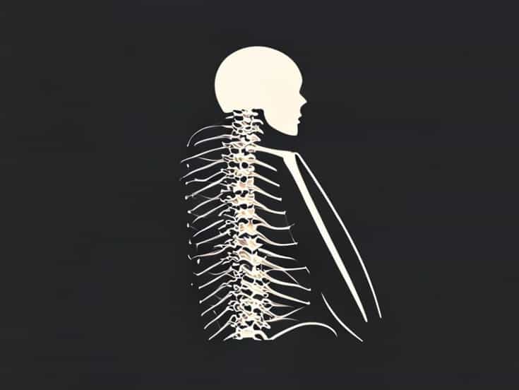
The spinal canal, also known as the vertebral canal, is a bony channel that extends from the base of the skull to the lower back. It is formed by the alignment of vertebral foramina, the openings in each vertebra, stacked vertically. This canal serves as a protective passageway for the spinal cord, spinal meninges, cerebrospinal fluid (CSF), and associated nerve roots. Its structure is designed to balance stability with flexibility, allowing movement while safeguarding the delicate neural tissues within.
General Structure
The spinal canal is tubular in shape and varies slightly in size and contour along different regions of the spine. It is wider in areas corresponding to regions with increased neural density, such as the cervical and lumbar enlargements, which accommodate nerve networks serving the limbs. The canal is narrower in the thoracic region due to the relatively smaller number of spinal nerve fibers passing through this segment. The walls of the spinal canal are formed by posterior portions of the vertebral bodies, the pedicles, laminae, and the posterior longitudinal ligament, creating a strong yet flexible protective compartment.
Regional Anatomy of the Spinal Canal
The spinal canal can be divided anatomically according to the spinal regions cervical, thoracic, lumbar, sacral, and coccygeal. Each region has unique characteristics that influence the size, shape, and function of the canal.
Cervical Region
The cervical spinal canal is located within the seven cervical vertebrae at the top of the vertebral column, beginning at the base of the skull and extending to the first thoracic vertebra. This portion of the canal is relatively wide and oval-shaped to accommodate the cervical enlargement of the spinal cord, which gives rise to the nerves supplying the upper limbs. The cervical canal provides a balance of protection and mobility, allowing significant neck movement without compromising spinal cord safety.
Thoracic Region
The thoracic spinal canal passes through the twelve thoracic vertebrae. It is narrower and more circular compared to the cervical region, reflecting the smaller cross-sectional diameter of the thoracic spinal cord. The canal is positioned posterior to the vertebral bodies and anterior to the laminae, with the pedicles forming the lateral walls. The thoracic canal’s design provides increased protection, which is particularly important due to the region’s limited flexibility and proximity to the ribcage and vital organs.
Lumbar Region
The lumbar spinal canal is located within the five lumbar vertebrae in the lower back. It is significantly wider than the thoracic canal, forming a more spacious compartment that accommodates the lumbar enlargement of the spinal cord and the emerging lumbar nerves supplying the lower limbs. The lumbar canal is posterior to the vertebral bodies, bordered laterally by the pedicles, and posteriorly by the laminae. Its dimensions are clinically significant because lumbar spinal stenosis can lead to compression of nerve roots, causing pain, numbness, or weakness in the lower extremities.
Sacral and Coccygeal Regions
The sacral canal is located within the sacrum, a triangular bone formed by the fusion of five sacral vertebrae. It continues the passage of spinal nerves into the sacral foramina, eventually contributing to the sacral and coccygeal plexuses. The coccygeal region, or tailbone, contains a small canal that usually houses the terminal filum of the spinal cord and connective tissue. These regions are narrower and more rigid, reflecting their role in weight-bearing and providing attachment points for ligaments and muscles.
Contents of the Spinal Canal
The spinal canal contains several crucial structures necessary for nervous system function
- Spinal CordThe central structure within the canal, extending from the brainstem to the lumbar region, transmitting sensory and motor signals.
- Spinal MeningesProtective membranes, including the dura mater, arachnoid mater, and pia mater, which surround the spinal cord and provide cushioning.
- Cerebrospinal Fluid (CSF)Fluid that circulates within the subarachnoid space, providing nourishment, shock absorption, and waste removal.
- Spinal Nerve RootsNerves emerging from the spinal cord that pass through the intervertebral foramina to innervate different parts of the body.
- Ligaments and Connective TissueStructures like the posterior longitudinal ligament, ligamentum flavum, and epidural fat, which stabilize and protect the spinal cord.
Clinical Significance of Spinal Canal Anatomy
Understanding the anatomic location of the spinal canal is essential in clinical practice. Knowledge of its size, shape, and contents is crucial for interpreting imaging studies, performing spinal surgery, administering epidural anesthesia, and diagnosing spinal pathologies such as stenosis, herniated discs, or tumors. Variations in canal dimensions can affect susceptibility to injury or nerve compression. For instance, congenital or acquired narrowing of the spinal canal, known as spinal stenosis, can lead to pain, numbness, and impaired motor function.
Procedural Implications
Medical procedures involving the spinal canal require precise anatomical knowledge
- Lumbar PuncturePerformed in the lumbar region, typically between L3-L4 or L4-L5, to access cerebrospinal fluid safely without damaging the spinal cord.
- Epidural AnesthesiaAdministered into the epidural space surrounding the spinal cord to provide regional anesthesia during surgeries or childbirth.
- Spinal SurgeryProcedures such as laminectomy, discectomy, or spinal fusion require detailed understanding of canal anatomy to avoid nerve injury.
The anatomic location of the spinal canal is central to the protection and function of the spinal cord and its associated structures. Extending from the base of the skull to the coccyx, the spinal canal varies in size and shape across cervical, thoracic, lumbar, sacral, and coccygeal regions to accommodate neural tissue and maintain flexibility. It houses the spinal cord, meninges, cerebrospinal fluid, and nerve roots, all protected by the bony vertebral column. Knowledge of its anatomy is essential in medicine for diagnosis, surgical intervention, and the management of spinal conditions. By understanding the spinal canal’s location, dimensions, and contents, healthcare professionals can ensure safe and effective care for patients with spinal or neurological concerns.