Ligamentum flavum hypertrophy is a condition commonly associated with spinal stenosis, and its evaluation through magnetic resonance imaging (MRI) has become a crucial tool in modern diagnostic medicine. This condition involves the thickening of the ligamentum flavum, a yellow elastic ligament that connects the laminae of adjacent vertebrae in the spine. Over time, degeneration, mechanical stress, and age-related changes can lead to hypertrophy, which may compress the spinal canal or nerve roots, resulting in pain, numbness, or weakness. MRI provides detailed visualization of soft tissue structures, making it the gold standard for identifying and assessing ligamentum flavum hypertrophy. Understanding its imaging features, clinical implications, and the significance of MRI findings is essential for healthcare providers managing spinal disorders, as well as for patients seeking clarity on their condition.
Understanding Ligamentum Flavum Hypertrophy
The ligamentum flavum is a critical structure in maintaining spinal stability and flexibility. Hypertrophy occurs when this ligament thickens beyond its normal range, often as a result of degenerative changes or repetitive stress. This thickening can encroach upon the spinal canal, contributing to lumbar or cervical spinal stenosis. While hypertrophy is commonly age-related, other factors such as chronic inflammation, trauma, or metabolic conditions can exacerbate the process. Patients with significant hypertrophy may experience symptoms like back pain, radiculopathy, claudication, or difficulty walking, emphasizing the importance of accurate diagnosis and timely intervention.
MRI as a Diagnostic Tool
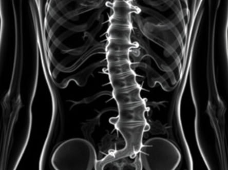
MRI is highly effective in evaluating ligamentum flavum hypertrophy because it provides excellent contrast between soft tissues and spinal structures. Unlike X-rays or CT scans, which primarily visualize bone, MRI allows clinicians to assess the thickness, composition, and impact of the ligament on the spinal canal. T1-weighted and T2-weighted sequences are typically used to highlight structural details. T1-weighted images provide information about anatomy and fat content, while T2-weighted images help visualize fluid, edema, or nerve compression, which may accompany ligamentum flavum hypertrophy.
MRI Findings in Ligamentum Flavum Hypertrophy
Characteristic MRI features of ligamentum flavum hypertrophy include thickened ligamentous tissue, often appearing as low-signal intensity on both T1- and T2-weighted images due to the dense collagen and elastin content. In cases of advanced hypertrophy, the ligament may bulge into the spinal canal, reducing space and potentially compressing the dura or nerve roots. Axial MRI slices are particularly useful for assessing the degree of spinal canal compromise, while sagittal views help evaluate the length and orientation of hypertrophic segments. MRI can also detect associated degenerative changes, such as facet joint arthropathy or disc bulging, which often coexist with ligamentum flavum hypertrophy.
Clinical Significance
Recognizing ligamentum flavum hypertrophy on MRI has important clinical implications. The degree of hypertrophy often correlates with the severity of symptoms, helping physicians decide on treatment strategies. Mild hypertrophy may be managed conservatively with physical therapy, anti-inflammatory medications, and lifestyle modifications, while severe hypertrophy causing significant spinal canal narrowing may necessitate surgical intervention. MRI findings provide a roadmap for procedures such as laminectomy or laminotomy, ensuring precise targeting of hypertrophic tissue while minimizing risks to surrounding structures.
Symptoms Linked to Ligamentum Flavum Hypertrophy
Patients with ligamentum flavum hypertrophy may present with a range of symptoms depending on the location and severity of spinal canal narrowing. Common presentations include
- Chronic lower back pain or neck pain
- Radiating pain along the legs or arms
- Numbness or tingling in extremities
- Weakness or difficulty in walking
- Neurogenic claudication, characterized by pain and discomfort after walking or standing for prolonged periods
These symptoms often prompt imaging studies, with MRI being the preferred modality due to its superior soft tissue visualization.
Factors Contributing to Hypertrophy
Several factors contribute to ligamentum flavum hypertrophy, and understanding these can aid in both prevention and treatment planning. Age-related degeneration is the most common factor, as the ligament loses elasticity and undergoes fibrosis over time. Repetitive mechanical stress, such as heavy lifting or prolonged poor posture, accelerates thickening. Inflammatory conditions, obesity, and genetic predisposition may also increase the likelihood of hypertrophy. MRI not only detects hypertrophy but can help identify these contributing factors indirectly by highlighting other degenerative changes within the spine.
Comparison with Other Imaging Modalities
While MRI is the gold standard, other imaging methods may complement the diagnosis of ligamentum flavum hypertrophy. CT scans can provide detailed information about bony structures and calcifications within the ligament, though soft tissue contrast is inferior. X-rays are generally limited to assessing vertebral alignment and degenerative disc disease but do not visualize the ligament itself. MRI remains superior for its ability to reveal both structural and pathological changes in soft tissues, making it essential for planning treatment strategies.
Management Based on MRI Findings
The management of ligamentum flavum hypertrophy largely depends on the severity of symptoms and the extent of canal compromise observed on MRI. Treatment options include conservative measures, minimally invasive procedures, and surgical interventions.
Conservative Treatments
- Physical therapy to improve flexibility and strengthen supporting muscles
- Anti-inflammatory medications to reduce pain and swelling
- Activity modification to avoid movements that exacerbate symptoms
- Weight management to reduce mechanical stress on the spine
Surgical Interventions
For patients with significant spinal canal narrowing or neurological deficits, surgery may be necessary. MRI helps guide surgical planning by accurately mapping the hypertrophic segments and assessing associated structures. Procedures such as laminectomy, laminotomy, or decompression surgery aim to remove or reduce hypertrophic ligament tissue, relieving pressure on neural elements and restoring spinal function.
Ligamentum flavum hypertrophy is a significant contributor to spinal stenosis, particularly in aging populations. MRI plays an indispensable role in diagnosing this condition, offering detailed visualization of soft tissue structures and associated degenerative changes. Recognizing the MRI features of hypertrophy allows clinicians to assess severity, correlate with patient symptoms, and plan effective treatment strategies, ranging from conservative therapy to surgical intervention. Early detection through MRI can prevent progression of neurological symptoms and improve overall patient outcomes. Patients experiencing back or neck pain, numbness, or difficulty walking should consult healthcare professionals for appropriate imaging and assessment. Understanding ligamentum flavum hypertrophy on MRI is essential for ensuring accurate diagnosis, informed decision-making, and optimal management of spinal disorders.
Word count ~1015