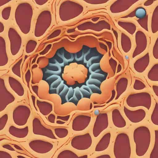
The epithelial lining of the proximal convoluted tubule (PCT) plays a crucial role in the kidney’s ability to filter blood and maintain homeostasis. As a fundamental component of the nephron, the PCT is responsible for the bulk reabsorption of water, electrolytes, glucose, amino acids, and other solutes from the filtrate back into the bloodstream. Understanding the structure and function of its epithelial lining provides insight into how the kidney efficiently manages fluid and electrolyte balance, as well as the removal of metabolic wastes. In this topic, we will explore the detailed anatomy of the PCT epithelium, its cellular components, functional mechanisms, and clinical significance.
Structure of the Proximal Convoluted Tubule
The proximal convoluted tubule is the first segment of the nephron following Bowman’s capsule. It is highly coiled, giving it a convoluted appearance, which increases its surface area for absorption. The epithelium lining the PCT is specialized to facilitate rapid reabsorption. Histologically, the PCT epithelium consists of a single layer of cuboidal cells with distinct features that optimize their function.
Characteristics of the Epithelial Lining
The epithelial cells of the PCT have several distinctive characteristics
- Brush BorderThe apical surface of PCT cells is covered with microvilli, forming a brush border. This dramatically increases the surface area available for reabsorption.
- Basolateral InfoldingsThe basal and lateral surfaces exhibit extensive infoldings that accommodate numerous mitochondria, providing energy for active transport mechanisms.
- Cytoplasmic ContentPCT cells contain abundant mitochondria, which supply ATP necessary for active transport of ions, glucose, and amino acids.
- Cell ShapeThe cells are cuboidal, with a central nucleus and dense cytoplasm, adapted for high metabolic activity and transport.
Functional Significance of the PCT Epithelium
The proximal convoluted tubule is responsible for reabsorbing approximately 65-70% of the glomerular filtrate. The epithelial lining facilitates both active and passive transport processes essential for maintaining body homeostasis.
Reabsorption of Solutes
The brush border microvilli are crucial for the efficient reabsorption of solutes. Key functions include
- Sodium ReabsorptionSodium ions are actively transported from the tubular lumen into the epithelial cells and subsequently into the bloodstream, creating osmotic gradients that drive water reabsorption.
- Glucose and Amino AcidsGlucose and amino acids are reabsorbed via sodium-dependent co-transporters. The high surface area of the brush border ensures that reabsorption is rapid and efficient.
- Chloride, Bicarbonate, and Other IonsChloride and bicarbonate ions are reabsorbed through both active and passive mechanisms, maintaining electrolyte balance and pH homeostasis.
Water Reabsorption
Water reabsorption in the PCT is largely driven by osmosis, which follows the reabsorption of solutes. The epithelial cells’ tight junctions allow water to pass selectively, ensuring that the majority of filtrate volume is reclaimed by the peritubular capillaries surrounding the PCT.
Secretion Functions
In addition to reabsorption, the PCT epithelial lining is involved in the secretion of certain substances from the blood into the tubular lumen. These include hydrogen ions, creatinine, and certain drugs or metabolites. The dense mitochondria in PCT cells supply the energy needed for these active transport processes.
Cellular Mechanisms and Transporters
The epithelial lining of the PCT contains specialized transporters and channels that facilitate reabsorption and secretion
- Na+/K+ ATPaseLocated on the basolateral membrane, this pump maintains low intracellular sodium levels, driving the active transport of sodium from the lumen.
- Sodium-Glucose Cotransporter (SGLT2)Located on the apical membrane, it reabsorbs glucose in conjunction with sodium ions.
- Na+/H+ ExchangerFacilitates sodium reabsorption and hydrogen ion secretion, playing a role in acid-base balance.
- Organic Anion and Cation TransportersInvolved in the secretion of metabolic wastes and xenobiotics into the tubular lumen.
Histological Appearance
Under a microscope, the proximal convoluted tubule’s epithelium appears as follows
- Cells with a prominent brush border creating a fuzzy apical surface.
- Large, centrally located nuclei within cuboidal cells.
- Abundant eosinophilic cytoplasm due to mitochondria concentration.
- Basolateral infoldings enhancing the surface area for interaction with peritubular capillaries.
The convoluted nature of the tubule and the dense cellular architecture highlight the high functional demand of this nephron segment.
Clinical Significance
Understanding the epithelial lining of the PCT is crucial in clinical medicine. Many kidney disorders and systemic diseases affect this segment of the nephron
- Acute Tubular NecrosisDamage to the proximal tubular epithelium, often due to ischemia or toxins, can impair reabsorption and lead to renal failure.
- Diabetic NephropathyHyperglycemia increases the workload on PCT cells, especially affecting glucose reabsorption via SGLT2 transporters.
- Drug ToxicityCertain drugs, such as aminoglycosides or chemotherapy agents, can selectively damage the PCT epithelium.
- Inherited DisordersConditions like Fanconi syndrome involve generalized dysfunction of proximal tubular reabsorption.
The epithelial lining of the proximal convoluted tubule is a highly specialized structure optimized for reabsorption and secretion. With its cuboidal cells, brush border, basolateral infoldings, and dense mitochondria, it enables the kidney to reclaim essential solutes and water while secreting metabolic wastes efficiently. Understanding this epithelium is critical not only for basic renal physiology but also for diagnosing and managing a variety of kidney disorders. The PCT epithelia demonstrate how structure is intricately linked to function, allowing the nephron to maintain the body’s fluid, electrolyte, and acid-base balance effectively.