The autonomic nervous system (ANS) is a critical part of the human body that controls involuntary physiological functions, including heart rate, digestion, respiratory rate, and glandular activity. Unlike the somatic nervous system, which regulates voluntary movements through skeletal muscles, the ANS operates automatically, ensuring that vital processes continue without conscious effort. At the heart of this system are the motor neurons of the autonomic nervous system, which transmit signals from the central nervous system to various target organs, coordinating responses that maintain homeostasis and adapt the body to changing internal and external environments. Understanding these neurons provides insight into how the body maintains balance and responds to stress, temperature changes, and other stimuli.
Overview of Motor Neurons in the Autonomic Nervous System
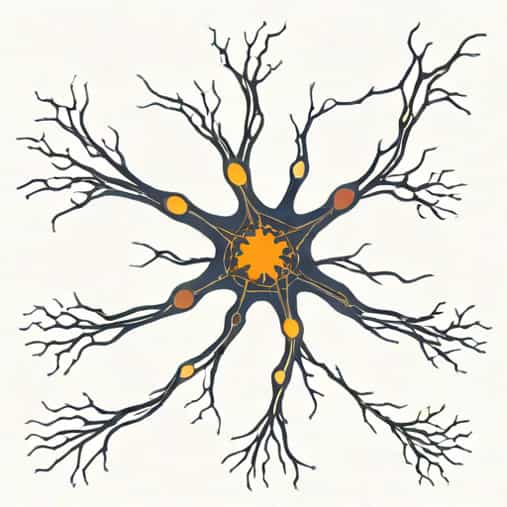
Motor neurons of the autonomic nervous system are specialized nerve cells that carry impulses from the brain and spinal cord to involuntary effectors, such as smooth muscles, cardiac muscles, and glands. These neurons are essential for controlling bodily functions that do not require conscious control, such as blood vessel constriction, heart rate modulation, and secretion of digestive enzymes. Unlike somatic motor neurons, which directly innervate skeletal muscles, ANS motor neurons usually operate through a two-neuron chain consisting of preganglionic and postganglionic neurons. ([h2]Motor Neurons of the ANS[/h2])
Preganglionic Neurons
Preganglionic neurons are the first in the two-neuron pathway of the autonomic system. Their cell bodies reside in the central nervous system, either in the brainstem or the spinal cord, depending on whether they belong to the sympathetic or parasympathetic division of the ANS. These neurons release neurotransmitters at autonomic ganglia, where they synapse with postganglionic neurons. The main neurotransmitter released by preganglionic neurons is acetylcholine (ACh), which binds to nicotinic receptors on postganglionic neurons, triggering the next stage of the signal transmission.
Postganglionic Neurons
Postganglionic neurons receive signals from preganglionic neurons at the autonomic ganglia and transmit them to target organs. The type of neurotransmitter released by postganglionic neurons depends on the division of the ANS
- Sympathetic Division Postganglionic neurons primarily release norepinephrine (noradrenaline), which prepares the body for fight or flight responses, such as increased heart rate and dilation of pupils.
- Parasympathetic Division Postganglionic neurons mainly release acetylcholine, promoting rest and digest activities, including stimulating digestive processes and slowing the heart rate.
These neurons are essential for modulating physiological responses appropriately to internal and external stimuli.
Divisions of the Autonomic Nervous System
The ANS is divided into the sympathetic and parasympathetic divisions, each with distinct motor neurons that regulate opposing physiological effects. Understanding these divisions is key to comprehending the function of motor neurons within the ANS.
Sympathetic Division
The sympathetic division originates primarily from the thoracolumbar region of the spinal cord. Preganglionic neurons are relatively short, synapsing with long postganglionic neurons in sympathetic ganglia located near the spinal column. This division is responsible for preparing the body to handle stressful or emergency situations, commonly referred to as the fight or flight response. Functions include
- Increasing heart rate and contractility
- Dilating pupils and bronchioles
- Diverting blood flow from the digestive system to skeletal muscles
- Stimulating sweat secretion
Parasympathetic Division
The parasympathetic division originates from the craniosacral regions, including the brainstem and the sacral spinal cord. Preganglionic neurons are long, synapsing with short postganglionic neurons located near or within the target organs. This division supports activities that conserve energy and promote bodily maintenance, known as rest and digest. Functions include
- Slowing heart rate
- Stimulating digestive enzyme secretion
- Promoting bladder and bowel function
- Constriction of pupils
Neurotransmitters and Receptors
The communication between motor neurons and target organs relies on neurotransmitters and receptors, which determine the effect of the ANS signal. Key neurotransmitters include
- Acetylcholine (ACh)Released by preganglionic neurons in both sympathetic and parasympathetic divisions and by postganglionic neurons in the parasympathetic division. It binds to nicotinic or muscarinic receptors depending on the target.
- Norepinephrine (Noradrenaline)Released by sympathetic postganglionic neurons, it binds to alpha and beta adrenergic receptors to induce various fight or flight responses.
- Other modulatorsCertain sympathetic neurons release epinephrine, dopamine, or other peptides in specialized tissues like adrenal medulla to amplify the response.
The specific combination of neurotransmitters and receptors allows motor neurons of the ANS to produce diverse and precise physiological effects tailored to the body’s needs.
Functional Significance
Motor neurons of the autonomic nervous system are essential for maintaining homeostasis and responding to environmental changes. Some examples include
- Cardiovascular Regulation Sympathetic neurons increase heart rate and contractility during stress, while parasympathetic neurons reduce heart rate during rest.
- Respiratory Control Sympathetic input dilates bronchioles, enhancing airflow, while parasympathetic input promotes normal airway tone.
- Digestive Function Parasympathetic neurons stimulate salivary secretion, peristalsis, and enzyme production, supporting digestion and nutrient absorption.
- Thermoregulation Sympathetic neurons control sweat glands and blood vessel diameter to regulate body temperature.
- Stress Response Sympathetic motor neurons prepare the body to respond quickly to threats by mobilizing energy stores and redirecting blood flow.
Clinical Relevance
Disorders of autonomic motor neurons can lead to serious health issues, including dysautonomia, hypertension, and irregular heart rates. Understanding how these neurons operate helps in diagnosing and treating conditions such as
- Autonomic neuropathy in diabetes
- Postural orthostatic tachycardia syndrome (POTS)
- Neurodegenerative disorders affecting autonomic function, like Parkinson’s disease
- Heart rate and blood pressure irregularities due to sympathetic or parasympathetic imbalance
Effective therapies often target neurotransmitters or receptors of the ANS to restore balance and normal physiological function.
The motor neurons of the autonomic nervous system play a pivotal role in controlling involuntary body functions, ensuring homeostasis, and enabling the body to respond appropriately to internal and external stimuli. Comprised of preganglionic and postganglionic neurons, these cells use neurotransmitters such as acetylcholine and norepinephrine to communicate with target organs. By modulating heart rate, digestion, respiration, and other critical processes, autonomic motor neurons support life-sustaining functions and adapt the body to varying demands. Understanding these neurons is essential for both medical science and clinical practice, offering insight into normal physiological regulation and the mechanisms behind autonomic disorders.