The human brain is an incredibly complex organ, and understanding how its different parts are organized helps us better understand how we think, move, and perceive the world. One common question in basic neuroscience and anatomy is how different regions of the brain relate to one another, especially when people learn that the frontal and occipital cortices are separated by specific structures. This topic may sound technical, but it can be explained in a clear and approachable way for general readers who are curious about how the brain is arranged and how its regions communicate.
Overview of the Frontal and Occipital Cortices
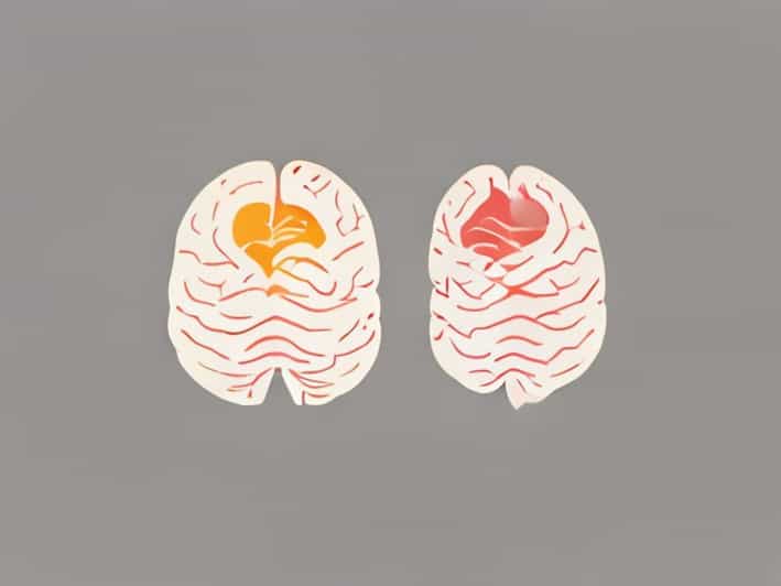
The cerebral cortex is the outer layer of the brain and is responsible for many higher-level functions such as thinking, planning, perception, and voluntary movement. It is divided into several regions called lobes, each with specialized roles. Among these, the frontal cortex and the occipital cortex are two of the most well-known and widely studied areas.
The frontal cortex, often referred to as the frontal lobe, is located at the front of the brain, just behind the forehead. It plays a major role in decision-making, problem-solving, emotional regulation, personality, and voluntary motor control. The occipital cortex, or occipital lobe, is located at the back of the brain and is primarily responsible for visual processing.
What Separates the Frontal and Occipital Cortices?
The frontal and occipital cortices are not directly adjacent to each other. Instead, they are separated by other cortical regions and anatomical landmarks. Most importantly, the parietal cortex and the temporal cortex lie between them, forming a continuous surface across the top, sides, and underside of the brain.
From an anatomical perspective, the frontal cortex is separated from the occipital cortex by distance rather than a single physical barrier. As you move from the front of the brain toward the back, you pass through the frontal cortex, then the parietal cortex, and finally reach the occipital cortex. This arrangement allows information to flow through multiple processing stages.
The Role of the Parietal Cortex
The parietal cortex sits between the frontal and occipital cortices along the upper part of the brain. It is involved in processing sensory information such as touch, temperature, pain, and spatial awareness. Because it connects with both frontal and occipital regions, it acts as an important bridge for integrating sensory input with action and decision-making.
For example, when you see an object using your occipital cortex and decide to reach for it using your frontal cortex, the parietal cortex helps coordinate the spatial and sensory information needed for that movement.
The Temporal Cortex as a Lateral Separator
On the sides of the brain, the temporal cortex also lies between the frontal and occipital cortices. The temporal lobe is involved in hearing, language comprehension, memory, and emotion. Although it does not sit directly on the top pathway between the frontal and occipital lobes, it plays a crucial role in linking perception, memory, and meaning.
This lateral separation shows that brain organization is not linear but three-dimensional. Different cortices are layered and folded in a way that maximizes surface area while maintaining efficient communication.
Major Sulci and Landmarks Involved
When discussing how the frontal and occipital cortices are separated, neuroscientists often refer to sulci, which are grooves or folds in the brain’s surface. These sulci help define boundaries between different lobes.
The Central Sulcus
The central sulcus is one of the most important landmarks in the brain. It separates the frontal cortex from the parietal cortex. While it does not directly separate the frontal and occipital cortices, it marks the transition from motor-related regions in the frontal lobe to sensory-related regions in the parietal lobe.
The Parieto-Occipital Sulcus
The parieto-occipital sulcus is a key structure that separates the parietal cortex from the occipital cortex. This sulcus is especially visible on the medial surface of the brain. Together with the central sulcus, it helps explain how the frontal and occipital cortices are separated through intermediate regions.
Functional Separation and Information Flow
Beyond physical separation, the frontal and occipital cortices are also functionally distinct. The occipital cortex processes raw visual input, such as shapes, colors, and movement. This information is then sent forward through a network of neural pathways to other regions, including the parietal and frontal cortices.
The frontal cortex uses this processed information to guide behavior, plan actions, and make decisions. This step-by-step flow shows why separation is beneficial. Each region specializes in certain tasks, reducing overload and increasing efficiency.
Visual Pathways Connecting Occipital and Frontal Areas
Although separated by other cortices, the frontal and occipital regions are strongly connected through neural pathways. Two major visual processing streams are often described
- The dorsal stream, which passes through the parietal cortex and helps with spatial awareness and movement
- The ventral stream, which passes through the temporal cortex and helps with object recognition
These pathways demonstrate how separation does not mean isolation. Instead, it allows complex collaboration between different brain regions.
Why This Separation Matters in Health and Disease
Understanding how the frontal and occipital cortices are separated is important in medicine and psychology. Damage to different regions leads to different symptoms. Injury to the occipital cortex can cause visual disturbances, while damage to the frontal cortex may affect personality, judgment, or movement.
Conditions such as stroke, traumatic brain injury, and tumors are often studied based on which cortical areas are involved. Knowing the spatial relationships between these regions helps clinicians diagnose problems and plan treatments more effectively.
Developmental and Evolutionary Perspectives
From a developmental point of view, the separation of cortical regions emerges as the brain grows and folds during early life. The increasing complexity of sulci and gyri allows for more specialized regions to develop. Evolutionarily, this organization reflects the increasing cognitive demands placed on the human brain.
As vision, motor control, and abstract thinking became more advanced, having distinct yet interconnected cortices provided a major advantage. The separation between frontal and occipital areas supports both specialization and integration.
The frontal and occipital cortices are separated not by a single structure, but by a combination of distance, intermediate cortical regions, and anatomical landmarks such as the parietal and temporal cortices and key sulci. This separation allows the brain to process information efficiently, moving from perception to action through well-organized pathways. By understanding this layout, general readers can gain a clearer picture of how the brain’s structure supports its remarkable range of functions.