The greater tubercle of the humerus is an important anatomical landmark that can be clearly identified on an x-ray of the shoulder. It plays a key role in muscle attachment and shoulder function, which makes it a frequent point of focus in diagnostic imaging. Whether a patient is experiencing shoulder pain, suspected fractures, or rotator cuff injuries, the greater tubercle often provides valuable information for healthcare professionals. Understanding how this structure appears on an x-ray, what it indicates, and why it matters in clinical diagnosis can help patients and medical students alike gain deeper insight into the shoulder joint’s complexity.
Anatomy of the Greater Tubercle of the Humerus
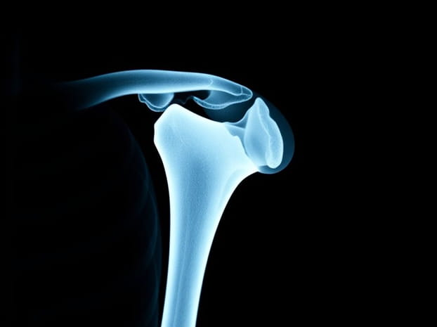
The humerus is the long bone of the upper arm, and the greater tubercle is one of its most prominent features. Located laterally on the proximal end of the humerus, the greater tubercle serves as the attachment site for several muscles of the rotator cuff. These muscles, including the supraspinatus, infraspinatus, and teres minor, play a crucial role in stabilizing and moving the shoulder joint.
Key Functions
- Provides leverage and attachment points for rotator cuff muscles.
- Assists in arm rotation and abduction movements.
- Acts as a protective structure for underlying tendons.
Appearance of the Greater Tubercle on X-Ray
On an x-ray, the greater tubercle appears as a rounded bony prominence on the lateral side of the humeral head. Its visibility depends on the projection of the x-ray, with certain angles making it clearer than others. Physicians often use anterior-posterior (AP), lateral, and specialized views to fully evaluate the greater tubercle.
Common Views Used
- AP ViewShows the greater tubercle as a lateral prominence adjacent to the humeral head.
- Lateral ViewHighlights its relation to the humeral shaft and surrounding structures.
- Oblique or Special ViewsSometimes used to better visualize subtle injuries or fractures.
Clinical Importance of the Greater Tubercle on X-Ray
The greater tubercle is a frequent site of injuries and degenerative changes, making its evaluation on x-rays particularly important. Physicians often assess this region when patients report shoulder trauma, chronic pain, or limited movement. Several conditions can be identified or suspected based on the appearance of the greater tubercle on x-ray.
Fractures of the Greater Tubercle
Fractures of the greater tubercle commonly occur due to falls, sports injuries, or high-impact accidents. On x-ray, these fractures may appear as small avulsions or more displaced breaks depending on the severity. Identifying the exact nature of the fracture is important for deciding whether conservative treatment or surgical intervention is needed.
Rotator Cuff Pathologies
Because rotator cuff tendons insert into the greater tubercle, abnormalities in this area may suggest tendon injuries. In cases of chronic shoulder pain, x-rays may show bony changes such as sclerosis, calcifications, or irregularities in the greater tubercle that point to rotator cuff disease.
Degenerative Changes
Over time, repetitive use of the shoulder joint can lead to degenerative changes visible in the greater tubercle area. These changes may include bone spurs or surface irregularities that indicate conditions such as osteoarthritis or chronic tendon impingement.
How Radiologists Interpret the Greater Tubercle
Radiologists carefully study the greater tubercle to evaluate bone alignment, shape, and density. Subtle differences in the outline or density may indicate fractures, soft tissue injuries, or chronic wear and tear. They also consider how the greater tubercle relates to other structures, such as the acromion and clavicle, to get a complete picture of shoulder health.
Key Points in Interpretation
- Symmetry between the greater tubercles of both shoulders.
- Presence of bone fragments suggesting fracture or avulsion.
- Signs of calcification related to tendon attachment issues.
- Overall joint space and alignment of the humeral head.
Greater Tubercle Injuries and Treatment
When an x-ray reveals abnormalities in the greater tubercle, treatment depends on the underlying condition. Fractures may require immobilization or surgery, while degenerative changes may be managed with physical therapy and lifestyle adjustments. The role of the x-ray is to guide these decisions by providing a clear picture of bone health.
Non-Surgical Management
In cases of minor fractures or tendon irritation, conservative management may include rest, anti-inflammatory medication, and physiotherapy. Strengthening exercises are often prescribed to support shoulder stability and prevent further injuries.
Surgical Options
More severe fractures of the greater tubercle or associated tendon injuries may require surgical repair. Procedures might involve fixation of bone fragments, tendon reattachment, or joint stabilization. Post-surgery, x-rays are also used to monitor healing progress.
Educational Importance for Medical Students
For students learning anatomy and radiology, the greater tubercle of the humerus provides a clear example of how bone landmarks are vital in both teaching and clinical practice. On an x-ray, identifying this structure helps learners develop a systematic approach to interpreting shoulder images. It also reinforces the connection between anatomy and clinical diagnosis.
Learning Tips
- Study the anatomy of the humerus before analyzing x-rays.
- Practice identifying the greater tubercle in different x-ray views.
- Compare normal and abnormal cases to sharpen diagnostic skills.
Greater Tubercle in Sports Medicine
Athletes often put significant strain on the shoulder, making the greater tubercle a vulnerable point for injury. In sports such as baseball, tennis, or swimming, repetitive overhead movements can lead to stress on the rotator cuff and greater tubercle. Sports physicians frequently rely on x-rays to assess damage and develop appropriate treatment plans.
Common Sports-Related Issues
- Avulsion fractures from sudden, forceful movements.
- Tendon inflammation leading to bone changes at the greater tubercle.
- Degenerative conditions from overuse.
Limitations of X-Ray in Evaluating the Greater Tubercle
Although x-rays provide valuable insights, they have limitations. Soft tissue injuries such as partial rotator cuff tears may not be fully visible. In such cases, MRI or ultrasound may be recommended for a more detailed view. However, x-rays remain the first-line imaging method because they are quick, accessible, and effective in detecting bone abnormalities.
The greater tubercle of the humerus on x-ray holds great importance in diagnosing shoulder conditions, guiding treatment, and teaching anatomy. As a prominent bone landmark, it reflects both the strength and vulnerability of the shoulder joint. From fractures and tendon injuries to degenerative changes, the greater tubercle provides vital clues for medical professionals. By understanding its appearance and significance on x-ray, patients, students, and clinicians can better appreciate the complex interactions between bone and muscle that allow the shoulder to function. In clinical practice, paying close attention to the greater tubercle ensures that no detail is overlooked in the pursuit of accurate diagnosis and effective treatment.